
Page 64 - GTM-3-1
P. 64
Global Translational Medicine Reproductive hormones and muscle in obesity
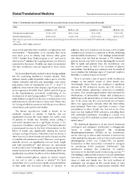
Table 5. Testosterone and estradiol levels in the musculus triceps surae tissues of the experimental animals
Index Male rats Female rats
Control (n = 13) HCD (n = 14) Control (n = 14) HCD (n = 13)
Testosterone (nmol/g tissue) 72.19 ± 15.95 68.91 ± 13.44 74.51 ± 9.96 71.47 ± 9.54
Estradiol (nmol/g tissue) 40.60 ± 10.52 41.18 ± 9.24 133.55 ± 11.82 146.83 ± 8.13 ##
Note: Data are presented as mean±standard deviation; statistically significant difference between the female HCD and female control groups at
## P < 0.01.
Abbreviation: HCD: High-calorie diet.
more slowly and have fewer metabolic complications with addition, fatty acid oxidation may increase until complete
overnutrition. Taraschenko et al. reported that males oxidation as β-oxidation is enhanced in obesity, indicating
17
were more prone to obesity than females when given mitochondrial dysfunction. Our findings demonstrated
27
HCD. Besides that, Chukijrungroat et al. and several that obese male rats had an increase in TP, cholesterol,
19
18
20
other studies validated the hepatoprotective role of female glucose, lactate, and LDH activity, implying the increased
reproductive hormones. Notably, our study demonstrated flow of lipids and glucose from the bloodstream into
that lipid metabolism was not impaired in obese female the muscle tissues, as well as the activation of glucose
rats. metabolism. This finding was consistent with the results of
recent studies that indicated signs of impaired metabolic
It is known that obesity can lead to male hypogonadism, flexibility in skeletal muscle in obesity. 27
but the underlying mechanism remains unclear. Diet-
induced obesity could reportedly reduce sperm motility, There is currently a lack of reports on the biochemical
relative testicular and testicular appendage mass ratios, changes in the muscle tissues of obese female rats.
and plasma levels of testosterone and LH in mice. In Interestingly, obese female rats displayed a significant
20
addition, obese rodents often display a significant decrease decrease in TP, cholesterol, lactate, and CK activity in
in the expression of GnRH, Kiss1, GpR54, and Ob-R genes the muscle tissues, indicating a reduction in metabolic
in the hypothalamus, potentially contributing to the processes. Our morphological analysis demonstrated the
development of male hypogonadism. 21,22 The results of our development of intracellular edema and inflammatory
study indicated a significant decrease in testosterone levels infiltration in the muscle tissue of obese male and female
and testosterone/estradiol ratio in obese male Wistar rats, rats. In the obese rats, the cross-sectional area of muscle
the finding of which is consistent with the previous clinical fibers was significantly reduced, while the total edema
observations and experimental studies. area increased. There was a significant increase in lipid
peroxidation in obese female rats, but not in the obese
The female reproductive health is known to be male rats that displayed SOD activation, providing them
markedly impaired in obesity. A high-fat diet can with antioxidant protection. According to a study, mice
significantly increase the body weight and earlier onset lacking Cu-Zn-SOD exhibited oxidative stress and loss of
of puberty in female rats. Likewise, estrus cycling is muscle mass. 28
commonly impaired due to a significant decrease in the
expression of ovulation-related genes. Our data indicated The functional importance of sex steroid hormones
23
a pronounced increase in the level of testosterone in the (androgens and estrogens) synthesized in extragonadal
8
blood of female rats, significantly altering the natural tissues is widely recognized. The circulating
estrogen-androgen balance. Polycystic ovary syndrome is dehydroepiandrosterone (DHEA) in the bloodstream
often observed in obese female rats and is characterized by can be taken up by tissues and converted to testosterone
elevated blood testosterone levels. Moreover, androgenic- when appropriate enzymes, such as 3-beta-hydroxysteroid
24
anabolic steroids can significantly increase body weight dehydrogenase (HSD) and 17-beta-HSD, are present.
and lipid peroxidation and decrease the antioxidant Testosterone is then irreversibly converted to estrogens
25
8
levels in female rats. Nonetheless, a significant increase in by cytochrome P-450 aromatase (P450arom). This
26
mechanism increases the production of anabolic
serum testosterone levels in females consuming HCD can hormones in the skeletal muscle of rats during exercise. 29,30
significantly exacerbate the effects of diet-induced obesity.
Our findings confirmed the presence of locally synthesized
Insulin-mediated glucose uptake in skeletal muscle hormones (i.e., testosterone and estradiol) in the muscle
27
in vivo and in vitro is reduced in obesity. For lipid tissue of male and female rats. The local synthesis of
metabolism, there is an increased uptake and accumulation androgens and estrogens in the muscle tissue of male
of fatty acids and decreased intensity of lipolysis. In rats presumably provides a compensatory anabolic effect
Volume 3 Issue 1 (2024) 6 https://doi.org/10.36922/gtm.2321