
Page 77 - GTM-3-4
P. 77
Global Translational Medicine Retrobulbar and sub-Tenon’s local anesthesia
enables quicker postoperative mobilization and patient and careful monitoring during the administration of
discharge. Three principal techniques – retrobulbar, retrobulbar anesthesia to minimize risks and ensure
2-5
peribulbar, and sub-Tenon’s block – are available for patient safety. 5,8-12 Brainstem anesthesia may occur due
delivering anesthetic agent around the cranial nerves to retrograde anesthetic flow from the ophthalmic artery
supplying the eye. The route of administration and the to the cerebral or internal carotid artery or due to dural
anatomical location of delivering the medication are the puncture around the optic nerve. This lethal complication,
main difference, but all aim to numb the nerves supplying while rare, can manifest as apnea, bradycardia, hypotension,
the ocular tissue. The difference in delivering medication and seizures. Surgical teams must be aware of preventive
may influence their efficacy and safety. 6,7 measures and equip the ophthalmic surgery room with
resuscitation equipment.
In retrobulbar and peribulbar anesthesia, a needle
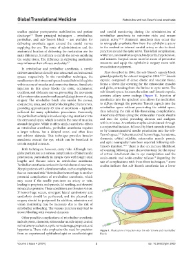
delivers anesthetics directly into intraconal and extraconal First described in 1884, the sub-Tenon’s capsule block
spaces, respectively. In the retrobulbar technique, the gained popularity for cataract surgery in 1994. 4,13,14 Tenon’s
needle enters the intraconal space, located behind the globe capsule, composed of dense elastic and vascular tissue,
within a cone of muscles and connective tissues. Anesthetic forms a sleeve-like covering for the extraocular muscles
injection in this space blocks the optic, oculomotor, and globe, extending from the limbus to optic nerve. The
trochlear, and abducens nerves, preventing the movement sub-Tenon’s space, between the sclera and Tenon’s capsule,
of the extraocular muscles and immobilizing the eye during contains ciliary nerve endings (Figure 1). Injection of
surgery. The retrobulbar block also numbs the cornea, anesthetics into the episcleral area allows the medication
conjunctiva, uvea, and sclera by blocking the ciliary nerves, to diffuse through the posterior Tenon’s capsule into the
providing approximately 45 min of anesthesia – typically retrobulbar space without penetrating the orbital space,
sufficient for many ophthalmic procedures. Conversely, thus reducing the risk of life-threatening complications.
the peribulbar technique involves injecting anesthetic into Anesthesia diffuses along the extraocular muscle sheaths
the extraconal space, which is outside the cone of muscles, and into the eyelid, providing akinesia and analgesia
around the globe. While it offers similar anesthetic effects within minutes. Anesthetics may be administered through
as retrobulbar anesthesia, peribulbar anesthesia requires a conjunctival incision, followed by blunt cannula infusion
a larger volume, has a delayed onset, and often does or by transconjunctival needle penetration into the sub-
4,15
not achieve akinesia. This technique provides broader Tenon’s space. Subconjunctival hemorrhage, hematoma,
anesthesia around the eye, which can be beneficial in chemosis, orbital cellulitis, extraocular muscle paresis,
certain surgical contexts. and optic neuropathy have been reported following sub-
Tenon’s injection. 16,17 There is also an increase likelihood
Both techniques, however, carry risks. Although rare, of vomiting following pars plana vitrectomy for the repair
globe perforation is a serious complication of blind needle of retinal detachment due to eye manipulation and the
penetration, particularly in myopic eyes with longer axial oculo-emetic and oculo-cardiac reflexes. Regarding the
18
lengths and thinner sclera in retrobulbar anesthesia. rate of complications with these three techniques, some
19
Peribulbar anesthesia carries similar risks but at a lower rate. studies indicate that sub-Tenon’s anesthesia has a lower
Myopic patients with scleral anomalies, such as staphyloma,
face an increased risk. Retrobulbar hemorrhage is another
8
potential complication of retrobulbar anesthesia, which
may occur if the needle punctures an artery or vein,
leading to proptosis, ecchymosis, lid swelling, and elevated
intraocular pressure. These conditions are threaten vision.
If hemorrhage occurs, emergent lateral canthotomy and
cantholysis should be performed, and the planned eye
surgery should be postponed. In addition, admission and
vision monitoring may be necessary due to the risk of
retrobulbar rebleeding. The venous puncture may lead to
slower bleeding with eventual chemosis.
Other possible complications of retrobulbar anesthesia
include ptosis, chemosis, extraocular muscle injury, central
retinal artery occlusion, optic nerve damage, proptosis, and
hypertonia. These risks emphasize the need for precision Figure 1. Illustration of injection sites for sub-Tenon’s and retrobulbar
from an experienced ophthalmologist or anesthesiologist blocks
Volume 3 Issue 4 (2024) 2 doi: 10.36922/gtm.3900