
Page 46 - GTM-4-1
P. 46
Global Translational Medicine Connective tissue harvest techniques
A B Table 1. A stepwise approach to single-incision subepithelial
connective tissue graft harvesting
Step Description
1 Identify the start and end points of the horizontal incision
based on anatomic limitations and the dimensions of the
required graft.
C D E 2 Connect the start and end points with an incision oriented
orthogonally to the surface of the palatal mucosa, extending to
the bone.
3 Reorient the scalpel to create a split-thickness incision in
the palatal tissue. Use the surface of the palatal mucosa and
the long axes of the maxillary teeth as anatomic landmarks.
Establish a primary flap thickness of approximately 1.5 mm.
4 Continue sharp dissection until the apicocoronal dimension of
the flap reaches 4 mm.
5 To reduce the risk of primary flap laceration, change the
F G scalpel angle by approximately 10°, directing the tip toward the
maxilla. Increase the depth of the sharp dissection until the
intended graft width is reached, respecting the apicocoronal
safety zone limitations.
6 Make vertical incisions and an apical horizontal incision to the
bone, outlining the graft.
7 Begin the graft harvest by reflecting the periosteum at the
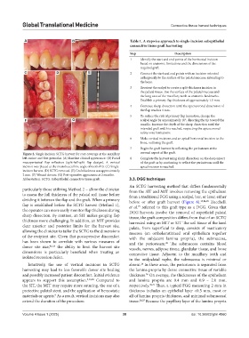
Figure 2. Single-incision SCTG harvest for root coverage at the maxillary coronal aspect of the graft.
left canine and first premolar. (A) Baseline clinical appearance. (B) Facial 8 Complete the harvest using sharp dissection on the deep aspect
mucoperiosteal flap reflection (split-full-split flap design). A vertical of the graft or by continuing to reflect the periosteum until the
incision was placed at the mesiobuccal line angle of tooth #13. (C) Single- apical incision is reached.
incision harvest. (D) SCTG removed. (E) Graft thickness was approximately
2 mm. (F) Wound closure. (G) Post-operative appearance at 2 months.
Abbreviation: SCTG: Subepithelial connective tissue graft. 3.3. DGG technique
An SCTG harvesting method that differs fundamentally
particularly those utilizing Method 2 – allow the clinician from the SIT and MIT involves removing the epithelium
to assess the full thickness of the palatal soft tissue before from a traditional FGG using a scalpel, bur, or laser, either
dividing it between the flap and the graft. When a primary before or after graft harvest (Figure 6). 15,24,25 Zucchelli
flap is established before the SCTG harvest (Method 1), et al. referred to this graft type as a DGG. Given that
15
the operator can more easily monitor flap thickness during DGG harvests involve the removal of superficial palatal
sharp dissection. By contrast, an SIT makes gauging flap tissue, the graft composition differs from that of an SCTG
thickness more challenging. In addition, an MIT provides harvested using an MIT or SIT. The soft tissue of the hard
clear anterior and posterior limits for the harvest site, palate, from superficial to deep, consists of masticatory
allowing the clinician to tailor the SCTG to the dimensions mucosa (an orthokeratinized oral epithelium together
of the recipient site. Given that postoperative discomfort with the subjacent lamina propria), the submucosa,
has been shown to correlate with various measures of and the periosteum. The submucosa contains blood
26
donor site size, 4,5,15 the ability to limit the harvest site vessels, nerves, adipose tissue, glandular tissue, and loose
dimensions is particularly beneficial when treating an connective tissue. Adjacent to the maxillary teeth and
isolated recession defect. in the midpalatal raphe, the submucosa is minimal or
Intuitively, the use of vertical incisions in SCTG absent. In these areas, the periosteum is separated from
26
harvesting may lead to less favorable donor site healing the lamina propria by dense connective tissue of variable
and possibly increased patient discomfort. Initial evidence thickness. On average, the thicknesses of the epithelium
26
appears to support this assumption. 9,16,23 Compared to and lamina propria are 0.4 mm and 0.9 – 2.0 mm,
the SIT, the MIT may require more suturing, the use of a respectively. 20,27 Thus, a typical FGG measuring 2 mm in
protective palatal stent, and the application of hemostatic thickness includes an epithelial layer <0.5 mm, most or
materials or agents. As a result, vertical incisions may also all of lamina propria thickness, and minimal submucosal
9
extend the duration of the procedure. tissue. 20,27 Because the papillary layer of the lamina propria
Volume 4 Issue 1 (2025) 38 doi: 10.36922/gtm.4860