
Page 49 - GTM-4-1
P. 49
Global Translational Medicine Connective tissue harvest techniques
A B C
D E F
G H I
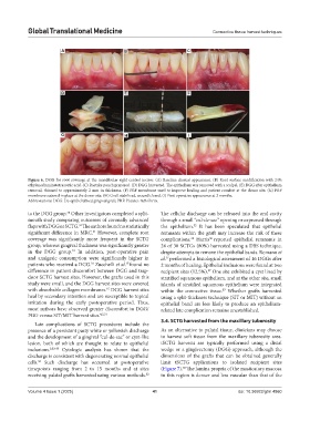
Figure 6. DGG for root coverage at the mandibular right central incisor. (A) Baseline clinical appearance. (B) Root surface modification with 24%
ethylenediaminetetraacetic acid. (C) Raetzke pouch prepared. (D) DGG harvested. The epithelium was removed with a scalpel. (E) DGG after epithelium
removal, thinned to approximately 2 mm in thickness. (F) PRF membrane used to improve healing and patient comfort at the donor site. (G) PRF
membrane sutured in place at the donor site. (H) Graft stabilized, wound closed. (I) Post-operative appearance at 2 months.
Abbreviations: DGG: De-epithelialized gingival graft; PRF: Platelet-rich fibrin.
to the DGG group. Other investigators completed a split- The cellular discharge can be released into the oral cavity
30
mouth study comparing outcomes of coronally advanced through a small “cul-de-sac” opening or expressed through
flaps with DGG or SCTG. The authors found no statistically the epithelium. It has been speculated that epithelial
31
32
31
significant difference in MRC. However, complete root remnants within the graft may increase the risk of these
coverage was significantly more frequent in the SCTG complications. Harris reported epithelial remnants in
36
32
group, whereas gingival thickness was significantly greater 24 of 30 SCTGs (80%) harvested using a DBS technique,
in the DGG group. In addition, post-operative pain despite attempts to remove the epithelial bands. Romano et
31
and analgesic consumption were significantly higher in al. performed a histological assessment of 16 DGGs after
37
15
patients who received a DGG. Zucchelli et al. found no 2 months of healing. Epithelial inclusions were found at two
31
difference in patient discomfort between DGG and trap- recipient sites (12.5%). One site exhibited a cyst lined by
37
door SCTG harvest sites. However, the grafts used in this stratified squamous epithelium, and at the other site, small
study were small, and the DGG harvest sites were covered islands of stratified squamous epithelium were integrated
with absorbable collagen membranes. DGG harvest sites within the connective tissue. Whether grafts harvested
15
37
heal by secondary intention and are susceptible to topical using a split-thickness technique (SIT or MIT) without an
irritation during the early postoperative period. Thus, epithelial band are less likely to produce an epithelium-
most authors have observed greater discomfort in DGG/ related late complication remains unestablished.
FGG versus SIT/MIT harvest sites. 4,5,31
3.4. SCTG harvested from the maxillary tuberosity
Late complications of SCTG procedures include the
presence of a persistent pasty white or yellowish discharge As an alternative to palatal tissue, clinicians may choose
and the development of a gingival “cul-de-sac” or cyst-like to harvest soft tissue from the maxillary tuberosity area.
lesion, both of which are thought to relate to epithelial tSCTG harvests are typically performed using a distal
inclusions. 2,32-35 Cytologic analysis has shown that the wedge or a gingivectomy (DGG) approach, although the
discharge is consistent with degenerating normal epithelial dimensions of the grafts that can be obtained generally
cells. Such discharge has occurred at postoperative limit tSCTG applications to isolated recipient sites
34
timepoints ranging from 2 to 15 months and at sites (Figure 7). The lamina propria of the masticatory mucosa
38
receiving palatal grafts harvested using various methods. in this region is denser and less vascular than that of the
32
Volume 4 Issue 1 (2025) 41 doi: 10.36922/gtm.4860