
Page 47 - GTM-4-1
P. 47
Global Translational Medicine Connective tissue harvest techniques
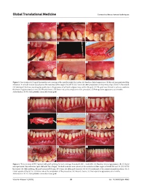
A B C
D E F
G H I
Figure 3. Two-incision SCTG graft harvest for root coverage at the maxillary right first molar. (A) Baseline clinical appearance. (B) Buccal mucoperiosteal flap
reflection. A vertical incision was placed at the mesiobuccal line angle of tooth #2. (C) Donor site after preparation of the primary flap. (D) SCTG harvested.
(E) Initial graft thickness was irregular, partly due to the presence of yellowish adipose tissue within the graft. (F) The graft was thinned to achieve a uniform
thickness of approximately 2 mm. (G) Wound closure. (H) Donor site at the completion of the procedure. (I) Post-operative appearance at 2 months.
Abbreviation: SCTG: Subepithelial connective tissue graft.
A B C
D E F
G H I
J K L
Figure 4. Three-incision SCTG harvest with graft splitting for root coverage from tooth #21 – tooth #29. (A) Baseline clinical appearance. (B, C) Facial
mucoperiosteal flap reflection (split-full-split flap design). Vertical incisions were placed at the mesiobuccal line angles of teeth #20 and 30. (D) SCTG
harvested. (E) After splitting, the graft measured 62 mm. (F) Donor site after graft removal. (G) SCTG positioned at the cementoenamel junctions. (H, I)
Distal aspects of the SCTG. (J) Donor site at the completion of the procedure. (K) Wound closure. (L) Post-operative appearance at 6 months.
Abbreviation: SCTG: Subepithelial connective tissue graft.
Volume 4 Issue 1 (2025) 39 doi: 10.36922/gtm.4860