
Page 46 - GTM-4-2
P. 46
Global Translational Medicine SPION for cancer theranostics
A B C
D E
F
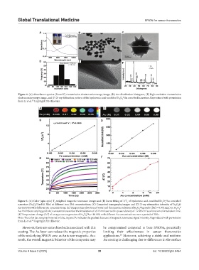
Figure 4. (A) Absorbance spectra (B and C) transmission electron microscopy image, (D) size distribution histogram, (E) high-resolution transmission
electron microscopy image, and (F) X-ray diffraction pattern of the hyaluronic acid-modified Fe O Au core/shell nanostars. Reproduced with permission
@
3
4
from Li et al. Copyright 2014 Elsevier.
35
A C
B D
E F
@
Figure 5. (A) Color (spin-spin) T -weighted magnetic resonance images and (B) linear fitting of 1/T of hyaluronic acid-modified Fe O Au core/shell
3
2
2
4
nanostars (Fe O Au-HA NSs) at different iron (Fe) concentrations. (C) Computed tomography images and (D) X-ray attenuation intensity of Fe O @
@
3
4
3
4
Au-HA NSs with different Au concentrations. (E) Temperature elevation of water and the aqueous solution of Fe O Ag seeds ([Fe] = 0.972 mm) or Fe O 4 @
@
3
4
3
2
Au-HA NSs at varying gold (Au) concentrations under the irradiation of a 915 nm laser with a power density of 1.2 W/cm as a function of irradiation time.
@
(F) Temperature change (ΔT) of an aqueous suspension of Fe O Au-HA NSs with different Au concentrations over a period of 300 s.
3
4
Note: The color bar, ranging from red to blue, in panel A indicates the gradual decrease of magnetic resonance signal intensity. Reproduced with permission
35
from Li et al. Copyright 2014 Elsevier.
However, there are some drawbacks associated with this be compromised compared to bare SPIONs, potentially
coating. The Au layer can reduce the magnetic properties limiting their effectiveness in cancer theranostics
of the underlying SPION core, as Au is non-magnetic. As a applications. Moreover, achieving a stable and uniform
30
result, the overall magnetic behavior of the composite may Au coating is challenging due to differences in the surface
Volume 4 Issue 2 (2025) 38 doi: 10.36922/gtm.8464