
Page 8 - IJB-1-1
P. 8
Smart hydrogels for 3D bioprinting
ser-based techniques. The latest progress in different 3. Bioprinting Techniques
smart hydrogel systems is presented while the chal-
lenges of printing these hydrogel systems are also 3D bioprinting can be generalized into three main
highlighted. Lastly, we present the future perspectives categories based on the technique being used, namely,
of smart hydrogels in 3D bioprinting. (i) Extrusion-based bioprinting techniques using me-
chanical forces to extrude the material through an
2. The Process and Flow of 3D Bioprinting orifice [32–33] , (ii) Inkjet-based techniques that eject
picoliter droplets of the material onto a substrate [34–37] ,
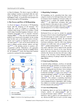
As shown in Figure 1, the process of 3D bioprinting
begins with the designing of 3D structures. This and (iii) Laser-based techniques where laser is used to
[38]
design file could be obtained by modelling via cure photopolymers or to induce material jetting .
computer-aided design (CAD) or by creating a virtual 3.1 Extrusion-based Bioprinting
model using biomedical imaging techniques, such as
computed tomography (CT) or magnetic resonance Mechanical forces are used to extrude the material
[32,33,39–41]
imaging (MRI) [22–24] . Next, the cells and the matrix are through an orifice in extrusion-based bioprinting .
prepared and transferred to the printer. In the printing Two different setups are usually employed in material
step, bio-inks such as the hydrogel matrix or biomo- extrusion-based bioprinting, namely the mechanical
lecules can be printed or dispensed into patterns for piston motor and pneumatic pumps. In these extru-
cell attachments [25] . Cells can be mixed with bio-inks sion-based techniques, materials with shear thinning
such as collagen, gelatin, matrigel, agarose, and algi- properties are ideally used, such as alginate [40–41] , ge-
nate [26–30] in the printing process to produce cell- latin [32] , gelatin methacrylamide [33] , etc. At the extru-
encapsulated scaffold. Alternatively, cell aggregates sion nozzle, high shear force is applied to the material
can be printed or patterned directly as building blocks while decreasing the viscosity of the shear thinning
for 3D structures [31] . Lastly, the bioprinted construct material for easy extrusion. Upon deposition onto the
needs to be incubated for tissue maturation. substrate, the decrease in shear rate will cause a sharp
increase in the viscosity of shear thinning materials,
enabling the printing of hydrogel strand with definite
shape fidelity.
3.2 Inkjet-based Bioprinting
In material jetting techniques, picoliters of materials
are dispensed onto the substrate, according to a prede-
termined design [34–36] . Structures are formed drop-by-
drop and subsequently layer-by-layer. Some examples
of material jetting techniques include, piezoelec-
tric/thermal inkjetting [34] , acoustic wave jetting [36] , and
electrohydrodynamic jetting [35] . In these techniques,
low viscosity of the material is an important
criterion [42] . This portrays a contrasting requirement
where the low viscosity material has to display suffi-
cient mechanical integrity immediately after printing.
One solution to overcome such a limitation is to cros-
slink the structure after printing [34,43,44] .
3.3 Laser-based Bioprinting
In bioprinting, lasers are used in two different tech-
nologies, namely vat polymerization [38] and la-
ser-induced forward transfer (LIFT) [45] . In vat poly-
merization, laser is used to cure photopolymers in a
Figure 1. Overview of the bioprinting procedure vat, whereas in LIFT, laser is used to provide energy
4 International Journal of Bioprinting (2015)–Volume 1, Issue 1