
Page 10 - IJB-1-1
P. 10
Smart hydrogels for 3D bioprinting
4.1 pH Responsive Hydrogel has been reported. However, further adaptation of ge-
lation condition is required to develop a printable ke-
pH responsive hydrogels are hydrogels that respond to ratin formulation.
environmental pH changes. They are made up of po-
lymeric backbones that can accept and/or donate pro-
tons during the change in pH [52,53] . Natural polymers
such as collagen and keratin are pH-responsive.
Collagen is the major component of extracellular
matrix protein. It consists three α chains which can
form triple helix. Collagen is a fibrous protein that
provides tensile strength to the extracellular matrix
(ECM). Type I Collagen is widely used for 2D sub- Figure 2. Keratin hydrogel (adapted from [13] )
strate coating and 3D scaffold. It can be dissolved in
acidic pH and form a gel at neutral pH and 37℃ [54] . The main limitation of pH responsive hydrogels is
In the work reported by Lee et al., a collagen hy- the non-physiological pH environment before or dur-
drogel precursor was used as a scaffold material for ing the gelation process. Cells cannot survive when
skin bioprinting. Nebulized sodium hydrogen carbo- being exposed to basic or acidic environment for
nate (NaHCO 3) vapor was applied onto the printed extended time during the bioprinting process.
collagen layer for gelation. Time lapse of one minute
was allowed to facilitate the phase transition of colla- 4.2 Temperature Responsive Hydrogel
gen to a gel, while cells were dispensed separately on Temperature responsive hydrogels can be further
top of the collagen gel [55] . classified as negative or positive temperature-
In another work, Park et al. bioprinted tissue-mi- responsive hydrogels. Positive temperature-responsive
metic structures composed of two compartments— hydrogels undergo sol-gel transition as the tempera-
cells-encapsulated hyaluronic acid (HA), and type I ture increases above critical solution temperature
collagen (Col-1) hydrogel for the cell migration study. (CST) [63–66] . Negative temperature-responsive hydro-
The hydrogel precursor’s solutions were first printed gels gel at the temperature below the CST [67,68] .
and the hydrogels were then cross-linked subsequently Censis et al. evaluated the suitability of a biode-
post-printing [56] . gradable, photopolymerizable and thermosensitive
3D Bi-Layered skin Tissue Formation Experiments A-B-A triblock copolymer hydrogel as a synthetic
with Collagen Hydrogel were reported by Koch et al. extracellular matrix for engineering tissues by means
In each cell–collagen layer, a mixture consisting of of three-dimensional fiber deposition. The polymer
collagen, sodium hydrogen carbonate (for neutralisa- was composed of poly(N-(2-hydroxypropyl) metha-
tion and gelification), and cells was used. The layer crylamide lactate) A-blocks, partly derivatized with
was then printed using Laser-assisted BioPrinting methacrylate groups, and hydrophilic poly(ethylene
(LaBP) [57] . glycol) B-blocks of a molecular weight of 10 kDa.
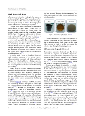
Keratins (Figure 2) are largely available, bioactive, Gels were obtained by thermal gelation and stabilized
and eco-friendly materials with gelation point at pH with additional chemical cross-links by photopolyme-
3.5. They also possess realistic potential as autologous rization of the methacrylate groups coupled to the
materials [58,59] . Keratins are intermediate filament polymer [69] .
proteins that are made up of intracellular cytoskele- Pluronic F127 is a favourite material for bioprinting
tons [60] . Despite not being ECM component proteins, due to its biocompatibility and gelling at the range of
11 out of 17 keratin subtypes contain cell binding se- 10 to 40℃ [70,71] . Chang et al. combined Pluronic F127
quence LDV (leu-asp-val), suggesting cell attachment with collagen type I as bioinks. Pluronic F127 has
on keratin materials through LDV recognition by α 4β 1 high viscosity at room temperature that can stay long
integrin [61] . Thus, keratin films and coatings have been enough for collagen gelation, while collage provides
used as cell culture substrates with demonstrated abil- necessary cell binding motifs for cell attachment.
ity to support cell attachment and growth [62] . Human Cells and fragments extracted from neovascular re-
dermal fibroblast encapsulated in 3D keratin hydrogel mained viable after printing using this method [47] .
6 International Journal of Bioprinting (2015)–Volume 1, Issue 1