
Page 100 - IJB-10-1
P. 100
International Journal of Bioprinting 3D bioprinting for musculoskeletal system
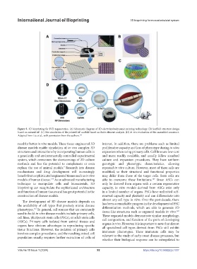
Figure 5. 3D bioprinting for IVD regeneration. (A) Schematic diagram of 3D electrohydrodynamic printing technology. (B) Scaffold structure design
based on natural AF. (C) The simulation of the printed AF scaffold based on finite element analysis. (D) In vivo evaluation of the assembled construct.
Adapted from Liu et al., with permission from the authors. 182
need for better in vitro models. These tissue-engineered 3D interest. In addition, there are problems such as limited
disease models enable simulation of in vivo complex 3D proliferative capacity and loss of phenotype during in vitro
structures and interactions by incorporating human cells in expansion when using primary cells. Cell lines are low-cost
a genetically and environmentally controlled experimental and more readily available, and usually follow standard
system, which overcomes the shortcomings of 2D culture culture and expansion procedures. They have uniform
methods and has the potential to complement or even genotypic and phenotypic characteristics, allowing
replace the use of animal models. Research into disease repeated in vitro culture. However, most of these cells are
7
mechanisms and drug development will increasingly modified, so their structural and functional properties
benefit from sophisticated engineered tissues such as in vitro may differ from those of the target cells. Stem cells are
models of human disease. As an advanced manufacturing able to overcome these limitations. Since ASCs can
187
179
technique to manipulate cells and biomaterials, 3D only be derived from organs with a certain regenerative
bioprinting can recapitulate the sophisticated architecture capacity, in vitro models derived from ASCs exist only
and function of human tissues and has great potential in the in a limited number of organs. PSCs have unlimited self-
construction of disease models. renewal capacity and plasticity and can differentiate into
The development of 3D disease models depends on almost any cell type in vitro. Over the past decade, there
the availability of cell types that precisely mimic disease has been a remarkable progress in the development of PSC
phenotypes. In general, cell sources that are commonly differentiation methods, which are able to generate 3D
181
183
used to build in vitro disease models include primary cells, tissue-like structures such as organoid models in vitro.
cell lines, pluripotent stem cells (PSCs), or adult stem cells These organoid models demonstrate similar morphology,
(ASCs). Primary cells isolated from animal tissues and cell composition, and function of the parts of developing
organs have obvious advantages in reproducing specific organs in vivo. However, it is important to note that almost
tissue functions. However, the isolation of primary cells all specialized cell types derived from PSCs still exhibit
involves complex procedures, and the resulting mixed cell immature phenotypes. These immature cells may be
population usually requires further extraction of cells of relevant to the study of early-onset disease processes, but
whether their biological response can be extrapolated to
Volume 10 Issue 1 (2024) 92 https://doi.org/10.36922/ijb.1037