
Page 311 - IJB-10-1
P. 311
International Journal of Bioprinting Low-cost quad-extrusion 3D bioprinting system
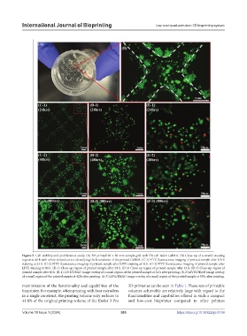
Figure 5. Cell viability and proliferation study. (A) 3D-printed 20 × 20 mm sample grid with 5% cell-laden GelMA. (B) Close-up of a strand crossing
region at 48 h with white-dotted curves identifying the boundaries of the printed GelMA. (C-1) FITC fluorescence imaging of printed sample after LIVE
staining at 24 h. (C-2) FITC fluorescence imaging of printed sample after LIVE staining at 42 h. (C-3) FITC fluorescence imaging of printed sample after
LIVE staining at 80 h. (D-1) Close-up region of printed sample after 24 h. (D-2) Close-up region of printed sample after 42 h. (D-3) Close-up region of
printed sample after 80 h. (E-1) LIVE/DEAD image overlay of a small region of the printed sample at 24 h after printing. (E-2) LIVE/DEAD image overlay
of a small region of the printed sample at 42 h after printing. (E-3) LIVE/DEAD image overlay of a small region of the printed sample at 80 h after printing.
maximization of the functionality and capabilities of the 3D printer as can be seen in Table 1. These sets of printable
bioprinter. For example, when printing with four extruders volumes achievable are relatively large with regard to the
in a single construct, the printing volume only reduces to functionalities and capabilities offered in such a compact
41.8% of the original printing volume of the Ender 3 Pro and low-cost bioprinter compared to other printers
Volume 10 Issue 1 (2024) 303 https://doi.org/10.36922/ijb.0159