
Page 328 - IJB-10-1
P. 328
International Journal of Bioprinting 3D-bioprinted meningioma model
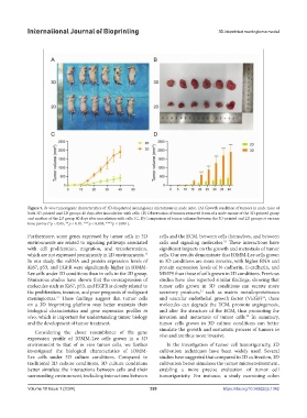
Figure 5. In vivo tumorigenic characteristics of 3D-bioprinted meningioma microtissue in nude mice. (A) Growth condition of tumors in nude mice of
both 3D-printed and 2D groups 40 days after inoculation with cells. (B) Observation of tumors removed from of a nude mouse of the 3D-printed group
and another of the 2D group 40 days after inoculation with cells. (C, D) Comparison of tumor volumes between the 3D-printed and 2D groups at various
time points (*p < 0.05, **p < 0.01, ***p < 0.005, ****p < 0.001).
Furthermore, some genes expressed by tumor cells in 3D cells and the ECM, between cells themselves, and between
environments are related to signaling pathways associated cells and signaling molecules. These interactions have
32
with cell proliferation, migration, and transformation, significant impacts on the growth and metastasis of tumor
which are not expressed prominently in 2D environments. cells. Our results demonstrate that IOMM-Lee cells grown
13
In our study, the mRNA and protein expression levels of in 3D conditions are more invasive, with higher RNA and
Ki67, p53, and EGFR were significantly higher in IOMM- protein expression levels of N-cadherin, E-cadherin, and
Lee cells under 3D conditions than in cells in the 2D group. MMP9 than those of cells grown in 2D conditions. Previous
Numerous studies have shown that the overexpression of studies have also reported similar findings, showing that
molecules such as Ki67, p53, and EGFR is closely related to tumor cells grown in 3D conditions can secrete more
the proliferation, invasion, and poor prognosis of malignant secretory products, such as matrix metalloproteinases
33
meningiomas. These findings suggest that tumor cells and vascular endothelial growth factor (VEGF) ; these
31
34
on a 3D bioprinting platform may better maintain their molecules can degrade the ECM, promote angiogenesis,
biological characteristics and gene expression profiles in and alter the structure of the ECM, thus promoting the
vivo, which is important for understanding tumor biology invasion and metastasis of tumor cells. In summary,
35
and the development of tumor treatment. tumor cells grown in 3D culture conditions can better
Considering the closer resemblance of the gene simulate the growth and metastatic process of tumors in
expression profile of IOMM-Lee cells grown in a 3D vivo and are thus more invasive.
environment to that of in vivo tumor cells, we further In the investigation of tumor cell tumorigenicity, 3D
investigated the biological characteristics of IOMM- cultivation techniques have been widely used. Several
Lee cells under 3D culture conditions. Compared to studies have suggested that compared to 2D cultivation, 3D
traditional 2D culture conditions, 3D culture conditions cultivation better simulates the tumor microenvironment,
better simulate the interactions between cells and their enabling a more precise evaluation of tumor cell
surrounding environment, including interactions between tumorigenicity. For instance, a study examining colon
Volume 10 Issue 1 (2024) 320 https://doi.org/10.36922/ijb.1342