
Page 327 - IJB-10-1
P. 327
International Journal of Bioprinting 3D-bioprinted meningioma model
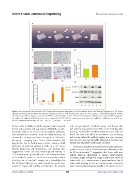
Figure 4. In vitro invasive characteristics of 3D-bioprinted meningioma microtissue. (A) Procedure for the in vitro Transwell invasion assay. (B) Tumor
cell invasion through the membrane was observed using a microscope in both the 3D-printed group and the 2D group, (C) followed by statistical analysis.
(D) The expression levels of genes associated with EMT, as determined by RT-qPCR. Normalization was performed using the expression of the target gene
(N-cadherin, E-cadherin, MMP9) relative to the expression of GAPDH. (E) The protein expression levels of N-cadherin, E-cadherin, and MMP9 were
detected using Western blotting (*p < 0.05, **p < 0.01, ***p < 0.005, ****p < 0.001).
of the coaxial scaffold provided a supportive environment this, we performed live/dead assays and found that
for the self-assembly and aggregation of IOMM-Lee cells. cell viability was greater than 90% on the first day after
Moreover, due to the absence of cell-scaffold adhesion, printing. Nevertheless, we observed an increase in the cell
cells were induced to interact with each other, forming cell death rate over time, which we attribute to the increasing
clusters that subsequently fused into spherical cell bodies, cell density within the scaffold, leading to a more compact
ultimately converging into a fibrous cellular network. We physical structure that limits the diffusion of nutrients and
hypothesize that the hollow nature of the coaxial scaffold oxygen and ultimately resulting in cell death.
facilitates unrestricted cellular growth in a 3D space, Previous studies have indicated that the gene expression
thereby promoting cell proliferation. Our findings also profile of tumor cells in 3D environments differs from that
suggest that IOMM-Lee cells exhibit high cell viability in in 2D environments, 28-29 suggesting that tumor cells may
a 3D environment, potentially due to the beneficial effects exhibit different phenotypes in distinct environments.
of the scaffold material on cell adhesion and growth, which Recently, a study showed that the gene expression profile of
may improve cell survival. However, we acknowledge that tumor cells in 3D environments is more similar to that of
the 3D bioprinting process and crosslinking agents may tumor cells growing in vivo and exhibits more in vivo-like
adversely impact cell viability and function. To address tumor characteristics than tumor cells in 2D environments.
30
Volume 10 Issue 1 (2024) 319 https://doi.org/10.36922/ijb.1342