
Page 322 - IJB-10-1
P. 322
International Journal of Bioprinting 3D-bioprinted meningioma model
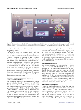
Figure 1. Schematic of the potential of the 3D-bioprinted meningioma model. (A) Schematic illustration of the coaxial bioprinting-based method and cell
microfiber structure. (B) Formation of cell fibers and cell balls from the inner axis. (C) The potential applications of 3D-printed tumor models.
2.2. Three-dimensional coaxial core-shell to a micropump containing the cell suspension with a flow
bioprinting method rate of 10 ml/h. The outer shell material was rapidly gelled
The concentric circle coaxial needle consists of a core in a crosslinking pool containing 3% (w/v) CaCl . After
2
channel with an inside diameter of approximately 0.38 mm approximately 5 min, the coaxial material was washed with
and a shell channel with an inside diameter of approximately 0.9% (w/v) NaCl solution five times and then transferred
1.1 mm, from which cells and materials can be extruded. to DMEM containing 10% (v/v) fetal bovine serum under
The coaxial needle was fixed on an iron frame, and its end sterile conditions. The cells were cultured in an incubator
was inserted into a crosslinking pool containing 3% (w/v) at 37°C with 5% (v/v) CO .
2
calcium chloride solution. The core and shell channels
were connected to injection pumps to control the flow rate. 2.4. Cell viability analysis
According to previously reported literature, the extrusion The viability of the cells was evaluated using a live/dead
19
speed of the shell was set at 10 ml/h, and the extrusion cell assay kit (KeyGEN BioTECH, Nanjing, China)
speed of the core was set at 35 ml/h. The needle was kept according to the manufacturer’s instructions. In brief,
facing downward and in the crosslinking pool throughout the 3D-bioprinted constructs were placed in a 10 ml
the process to ensure stable flow rates. After the extrusion mixture of 1× phosphate-buffered saline (PBS) containing
process was complete, the 3D cell fibers obtained were 8 μM propidium iodide (PI) and 2 μM calcein-AM and
removed and thoroughly cleaned. incubated in the dark at room temperature for 10 min.
Subsequently, the 3D-bioprinted constructs were washed
2.3. Three-dimensional meningioma model three times with 1× PBS, and images of the green (live) and
establishment and culture red (dead) fluorescence were captured using a fluorescence
For preparation of the outer shell material, sodium alginate microscope (Eclipse Ti2-U, Nikon, Japan).
powder (Aladdin, China) was dissolved in 0.9% (w/v)
NaCl to form a 1.5% (w/v) sodium alginate solution, which 2.5. Cell proliferation assay
was subsequently sterilized by high-temperature and high- For analysis of cell proliferation, an Alamar Blue assay was
pressure treatment. A 3% (w/v) CaCl solution (Sigma- conducted according to the manufacturer’s instructions
2
Aldrich, Shanghai, China) was used for crosslinking and (Yeasen, China). In brief, 3D-bioprinted constructs,
was also sterilized. IOMM-Lee cells were harvested from approximately 5 cm in length, were prepared in each well
2D culture dishes and suspended in DMEM at a density of a 6-well plate. Next, 2 ml of a 1:10 mixture of Alamar
of 5 × 10 cells/ml. The outer shell was connected to a Blue and cell culture medium was added to each well of
7
micropump containing the sodium alginate solution with the 6-well plate and incubated at 37°C in the dark for 4
a flow rate of 35 ml/h, while the inner core was connected h for detection. After Alamar Blue incubation, 100 μl of
Volume 10 Issue 1 (2024) 314 https://doi.org/10.36922/ijb.1342