
Page 325 - IJB-10-1
P. 325
International Journal of Bioprinting 3D-bioprinted meningioma model
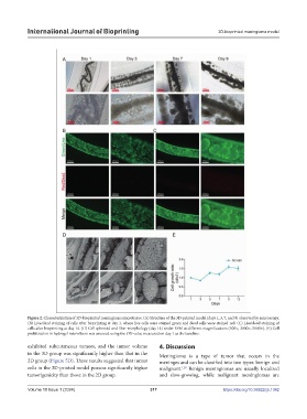
Figure 2. Characterization of 3D-bioprinted meningioma microtissue. (A) Structure of the 3D-printed model (days 1, 3, 7, and 9) observed by microscopy.
(B) Live/dead staining of cells after bioprinting at day 1, where live cells were stained green and dead cells were stained red. (C) Live/dead staining of
cells after bioprinting at day 14. (D) Cell spheroid and fiber morphology (day 14) under SEM at different magnifications (500×, 2000×, 5000×). (E) Cell
proliferation in hydrogel microfibers was assessed using the OD value measured on day 1 as the baseline.
exhibited subcutaneous tumors, and the tumor volume 4. Discussion
in the 3D group was significantly higher than that in the Meningioma is a type of tumor that occurs in the
2D group (Figure 5D). These results suggested that tumor meninges and can be classified into two types: benign and
cells in the 3D-printed model possess significantly higher malignant. Benign meningiomas are usually localized
1,20
tumorigenicity than those in the 2D group. and slow-growing, while malignant meningiomas are
Volume 10 Issue 1 (2024) 317 https://doi.org/10.36922/ijb.1342