Page 488 - IJB-10-1
P. 488
International Journal of Bioprinting                             Endothelial monolayer formation on scaffolds


            2.3. Scaffold coating with fibrin                  described 19,20  to enable cell alignment with the flow. A
            Coating was performed by application of human fibrinogen   schematic of the conditions in the slide is depicted in
            (10  mg  ml   in  PBS;  Sigma-Aldrich,  St.  Louis,  USA)   Figure S2  (Supplementary File). After cultivation, cells
                     -1
            solution for 10 s to the scaffold before crosslinking using   were  fixed,  and fluorescence  staining  was performed.
            5 µl of human thrombin (diluted to 5 U ml  in PBS; Sigma-  Therefore, scaffolds were incubated with 0.1%  Triton-X
                                             -1
            Aldrich,  St.  Louis,  USA)  to  each  scaffold.  Crosslinked,   (Sigma-Aldrich,  USA)  for  1  h.  Next,  the  scaffolds  were
            coated scaffolds were immersed repeatedly in fibrinogen   stained with phalloidin (Phalloidin-iFluor  555, Abcam
                                                                                                 TM
            solution for 10 s followed by 1 h of incubation at room   PLC, UK) for 90 min and washed again six times with PBS.
            temperature. The initial amount of thrombin was sufficient   Incubation with 300 nM DAPI solution (Sigma-Aldrich,
            to accomplish the crosslinking after further coating steps.  USA) was carried out for 30  min. Subsequently, VE-
                                                               cadherin staining was performed by overnight incubation
            2.4. Characterization of the fibrin layer          with an anti-VE cadherin antibody (rabbit origin, Abcam
            The fibrin layer, resulting from repeated scaffold coating,   plc., UK). Antibody staining was performed by incubation
            was visualized by immunostaining. Therefore, scaffolds   for 2 h with conjugated antibody to the first antibody (goat
            were fixed with 4 wt% paraformaldehyde for 30 min and   anti-rabbit Alexa Fluor® 488; Abcam PLC, Cambridge, UK).
            then washed three times with PBS. Scaffolds were incubated   Scaffolds were washed thrice with PBS before microscopy.
            with an anti-fibrinogen antibody (anti-fibrinogen alpha   KLF2 staining was carried out by overnight incubation at
            chain antibody; Abcam PLC, Cambridge, UK) overnight   8°C in PBS containing an anti-KLF2 antibody in a dilution
            at 4°C before incubation with a fluorophore-linked anti-  of 1: 150 (mouse origin; Abcam PLC, UK) after fixation
            fibrinogen antibody (Goat Anti-Rabbit IgG H&L Alexa   and incubation with Triton-X. Antibody staining was
            Fluor® 555, Abcam PLC, Cambridge, UK) for 1 h. Stained   visualized by incubation for 2 h with a label-conjugated
            scaffolds were evaluated microscopically to evaluate   second antibody (goat  anti-mouse  Alexa  Fluor®  555,
            fibrin layer  thickness and pore size. Image acquisition   Abcam PLC, Cambridge, UK).
            and analysis were performed using an Olympus IX83
            fluorescence microscope in combination with the software   Images  of  cells  and  scaffolds  were  captured  using
            cellSens dimensions (Olympus K.K., Shinjuku, Japan).   an Olympus IX-83 inverted microscope. Multichannel
            Z-stacking was used to increase the depth of field.  fluorescence images were taken with Z-stacking
                                                               algorithms to expand the depth of field. In the case of KLF2
            2.5. Static and dynamic cultivation of HUVECs on   immunostaining, image acquisition was carried out with
            MEW and MES scaffolds                              the same exposure time, gain, and contrast for all images
            HUVECs (PromoCell GmbH, Heidelberg, Germany)       to obtain comparable results.
            were used in all static and dynamic experiments. HUVEC
            suspension with a density of 250,000  cells  cm  in 20  µl   The axis rotation and the roundness of the HUVECs
                                                  -2
            of EGM2 was added to the scaffolds, which were then   were analyzed based on the cell shape visualized through
            incubated for 1 h to promote cell adhesion before surplus   the  VE-cadherin  staining.  In  detail,  axis  rotation  and
            cell  culture  medium  was  added.  Cultivations  were   roundness were calculated after image processing using
            performed at 37°C and 5%  pCO . EGM2 (PromoCell    Fiji as described in  Figures S3 and  S4  (Supplementary
                                        2
            GmbH, Heidelberg, Germany), supplemented with      File). By performing contrast enhancements, threshold
            additional 8% fetal bovine serum (Sigma-Aldrich, USA)   adjustments, and by using filters such as “find edges” and
            and 0.5% gentamicin (10 mg ml , Sigma-Aldrich, St.   “unsharp mask,” a binary mask for recognition of structural
                                        -1
            Louis, USA), was used for all experiments. The medium   features was created. Ten  pictures were analyzed out of
            was changed every 2 days during the static cultivation   three separate scaffolds from separate cultivations with cell
            experiments.                                       numbers ranging from 1265 to 2812 cells per experimental
                                                               setup (median = 1856 cells), depending on the conditions
               Dynamic cell culture experiments on scaffolds were   of the cultivation.
            performed using coated µ-slides I Luer 3D (ibidi GmbH,
            Gräfelfing,  Germany). Scaffolds were  cultivated  for  7   For the assessment of altered cell roundness under the
            days under static conditions and transferred into the   influence of flow, the cell area (A) and the longest axis (L)
            molds of the µ-slide. The slides were connected to the   were measured. Cell elongation was evaluated using the
            perfusion system (ibidi GmbH, Gräfelfing, Germany),   parameter “roundness” (0 = straight line, 1 = perfect circle)
                                                                               21
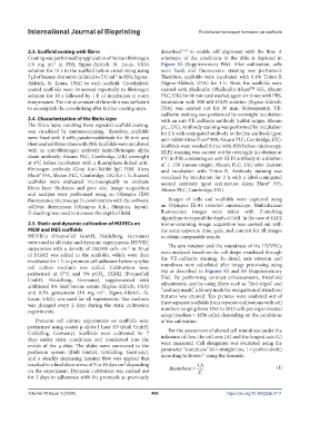
            and a steadily increasing laminar flow was applied that   according to Brown  using the formula:
            resulted in a final shear stress of 5 or 10 dyn cm² depending    4 A                           (I)
            on the experiment. Dynamic cultivation was carried out   Roundness =  L 2
            for 3 days in adherence with the protocols as previously


            Volume 10 Issue 1 (2024)                       480                          https://doi.org/10.36922/ijb.1111

