
Page 490 - IJB-10-1
P. 490
International Journal of Bioprinting Endothelial monolayer formation on scaffolds
3.2. Impact of fibrinogen coating on confluent 3.3. Determination of optimal pore size in MEW for
endothelium formation confluent endothelial formation
To achieve an even and complete fibrin coating on the The pore size of scaffolds is an important property to enable
MEW scaffolds, the optimal number of immersion steps gapless tissue formation. Here, an interfiber distance of
and fibrinogen concentration used in the coating process 40, 60, 80, 100, and 120 µm was evaluated for confluent
were determined to achieve confluent endothelial cell endothelial formation (Figure 3A and B). It is noted that
growth. Here, we used PCL grids with fiber spacing of 58.6 ± 31.07% of all 120 µm-sized pores were overgrown
80 µm as a proof of principle, but we assume that the fibrin by cells, while the remaining 41.4 % were not overgrown by
amount accumulated on the scaffold would not differ with cells, resulting in gaps in the cell layer as seen in Figure 3C.
a narrower fiber spacing, and the fibrin would be even The percentage of areas with overgrown pores increased
more evenly distributed with a narrower fiber spacing. with decreasing interfiber distance, reaching a maximum
of 96.2 ± 1.03% overgrown pores at an interfiber spacing of
With all tested fibrinogen concentrations, a single
coating step resulted in irregular cell seeding and cell 40 µm. The percentage of overgrown pores for 60, 80, and
growth over the pores (Figure 2A and B), whereas repeated 100 µm-diameter pores was 90.9 ± 4.29%, 90.6 ± 5.03%,
coating steps led to confluent endothelial layers (Figure 2C– and 66.5 ± 4.52% (n = 3), respectively. To optimize cell
F). More than two coating steps and concentrations higher layer formation on the scaffold, the interfiber distance
-1
than 10 mg ml of fibrinogen led to superfluous fibrin on was set to 40 µm for all future experiments. Under these
the scaffold and chaotic endothelial cell layers. Continuous conditions, we observed the smallest amount of gaps in the
endothelial layers are necessary to avoid cell detachment cell layer and the best reproducibility (as represented by
during dynamic cultivation. They are a prerequisite the smallest standard deviation).
for exposure of the seeded cells to uniform shear stress The use of non-square pores did not affect cell
during dynamic cultivation. Therefore, further cell culture orientation. Regardless of the pore dimension, cells
experiments were performed with two coating steps using displayed a cobblestone-like phenotype under static
10 mg ml fibrinogen to achieve confluent monolayers. conditions (Figure 3D).
1
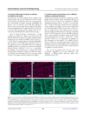
Figure 2. Culture of HUVECs on MEW/FDM scaffolds handled with different fibrin coating procedures. The procedure involved testing with one, two, or
three coating steps with 10, 15, and 20 mg ml fibrinogen or without any fibrinogen. Each panel shows HUVECs cultured on MEW/FDM scaffolds handled
1
with different fibrinogen coating procedures: (A) no coating, (B) 1 × 20 mg ml , (C) 2 × 10 mg ml , (D) 2 × 20 mg ml , (E) 3 × 10 mg ml , and (F) 3 ×
-1
-1
-1
-1
20 mg ml . Polymerization of fibrinogen to fibrin was facilitated by 5 µl thrombin solution (5 U ml ). Cells were seeded with a density of 250,000 cm and
-1
2
1
cultivated for 7 days on MEW scaffolds with 80 µm fiber spacing. Cell nuclei are labeled in blue, actin stained in red, and VE-cadherin in green. For each
condition, three separate scaffolds were evaluated (n = 3).
Volume 10 Issue 1 (2024) 482 https://doi.org/10.36922/ijb.1111