
Page 494 - IJB-10-1
P. 494
International Journal of Bioprinting Endothelial monolayer formation on scaffolds
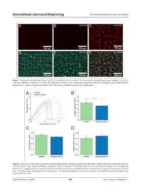
Figure 7. Expression of Krüppel-like factor 2 (KLF2) of endothelial cells on scaffolds. Cells were either cultivated under static conditions (A and D),
-2
-2
5 dyn cm (B and E), or 10 dyn cm (C and F). KLF2 was stained in red (A–C); combined staining with VE-cadherin and DAPI was also performed (D–F).
Exposure time, contrast, and gain were fixed for KLF2 microscopic evaluation to obtain comparable results.
Figure 8. Comparison of mechanical properties of seeded and unseeded scaffolds evaluated by tensile testing. Samples were either seeded with HUVECs
and cultivated for 7 days (seeded scaffold) or incubated in PBS for 7 days at 37°C (scaffold). Stress–strain curves of specimen tested in tensile testing are
shown in (A). Young’s modulus (B), tensile strain (C), and tensile stress (D) were evaluated from the measured data. The extension rate was set to 20 mm
min . Error bars indicate the standard error of the mean (n = 5). Statistical significance was examined using two-way ANOVA, and statistical significance
-1
is indicated by *p < 0.05.
Volume 10 Issue 1 (2024) 486 https://doi.org/10.36922/ijb.1111