
Page 88 - IJB-10-1
P. 88
International Journal of Bioprinting 3D bioprinting for musculoskeletal system
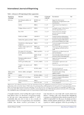
Table 1. Advances in 3D bioprinting for bone regeneration
Bioprinting Materials Cell type Cell density Key outcomes Ref.
technology (cells/mL)
Extrusion HA, fibrinogen, gelatin, and HUVECs and 1 × 10 7 Supported robust vascular 53
glycerol BMSCs development and higher levels of new
bone formation
GelMA BMSCs 5 × 10 6 Promoted new bone formation in 54
vivo
Collagen, chitosan, and β-GP BMSCs 5 × 10 7 Facilitated osteogenic differentiation 55
and bone regeneration in vivo
Bone ECM ADSCs 1.2 × 10 7 Promoted new bone formation 56
and more competent vascular
development
HAMA and GelMA C3H10T1/2 1 × 10 7 Promoted osteoblast differentiation 57
and induced ectopic bone formation
GelMA, PEG, gelatin, and MSN BMSCs 1 × 10 7 Promoted osteogenic differentiation 58
and accelerated diabetic bone repair
ACuMBGNs, oxidized alginate, BMSCs 1 × 10 6 Promoted osteogenic differentiation 59
and gelatin and angiogenesis
HAMA, GelMA, alginate, and BMSCs and 2 × 10 6 Promoted the M2-type polarization 60
graphene oxide macrophages of macrophages and promoted bone
repair
HA, gelatin, PCL, fibrinogen, BMSCs and EPCs 1.5 × 10 7 Promoted the new blood vessels and 61
PF-127, glycerol, and thrombin new bone formation
GelMA, HERS cells and 1 × 10 6 Generated mineralization texture and 52
DPCs promoted alveolar bone regeneration
Fibrinogen, gelatin, glycerol, BMSCs 5 × 10 6 Supported bone formation and 62
HA, and PCL vascularization
GelMA, gum methacrylate HUVECs, BMSCs 2 × 10 6 Promoted bone regeneration and 63
angiogenesis
Graphene oxide, alginate, and BMSCs 5 × 10 7 Promoted osteogenic differentiation 66
gelatin
Bone ECM HUVECs, MSCs 1 × 10 7 Led to the formation of 65
interconnected vascular networks
Robotic in situ PEGDA, GelMA, and alginate MC3T3-E1 cells - Promoted the repair of long 66
extrusion segmental defects
VBP GelMA HUVECs, BMSCs 3 × 10 6 Promoted osteogenic differentiation 67
LAB BioRoot RCS® and collagen Stromal cells 7 × 10 7 Promoted osteogenic differentiation 68
and bone formation
DLP GelMA and dextran BMSCs - Promoted bone regeneration in vivo 69
SilMA MC3T3-E1 cells 2 × 10 6 Drove osteogenesis 70
Abbreviations: VBP: volumetric bioprinting, LAB: laser-assisted bioprinting, DLP: digital light processing, HA: hyaluronic acid, GelMA: gelatin
methacrylate, ECM: extracellular matrix, HAMA: hyaluronic acid methacrylate, MSN: mesoporous silica nanoparticle, PCL: polycaprolactone, PEGDA,
SilMA: silk fibroin methacrylate, β-GP: β-glycerophosphate, PF-127: Pluronic F-127, HUVECs: human umbilical vein endothelial cells, BMSCs: bone
marrow stem cells, ADSCs: adipose-derived stem cells, EPCs: endothelial progenitor cells, HERS: Hertwig’s epithelial root sheath, DPCs: dental papilla
cells, ACuMBGNs: amine-functionalized copper (Cu)-doped mesoporous bioactive glass nanoparticles
of vascular networks, which facilitate the repair of critical bone formation in a rat model with cranial critical-sized
bone defects. Shen et al. developed a bioprinting strategy defects. Another study used intraoperative bioprinting
54
to fabricate bone tissue-engineered scaffolds in which to prepare a scaffold that enabled simultaneous delivery of
endothelial cells were able to form in situ networks of pPDGF-B and pBMP-2 for the repair of critical-sized bone
blood vessels. The in vivo bioprinted in situ vascularized defects. Platelet-derived growth factor (PDGF) has been
54
scaffolds have shown excellent performance in new reported to exhibit angiogenic effects by promoting the
Volume 10 Issue 1 (2024) 80 https://doi.org/10.36922/ijb.1037