
Page 89 - IJB-10-1
P. 89
International Journal of Bioprinting 3D bioprinting for musculoskeletal system
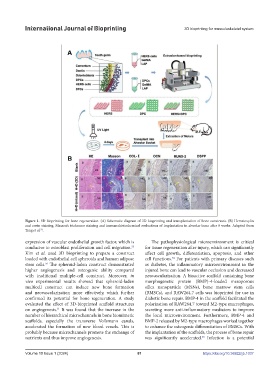
Figure 1. 3D bioprinting for bone regeneration. (A) Schematic diagram of 3D bioprinting and transplantation of bone constructs. (B) Hematoxylin
and eosin staining, Masson’s trichrome staining and immunohistochemical evaluations of implantation in alveolar bone after 8 weeks. Adapted from
Tang et al. .
54
expression of vascular endothelial growth factor, which is The pathophysiological microenvironment is critical
conducive to osteoblast proliferation and cell migration. for tissue regeneration after injury, which can significantly
55
Kim et al. used 3D bioprinting to prepare a construct affect cell growth, differentiation, apoptosis, and other
loaded with endothelial cell spheroids and human adipose cell functions. For patients with primary diseases such
74
stem cells. The spheroid-laden construct demonstrated as diabetes, the inflammatory microenvironment in the
56
higher angiogenesis and osteogenic ability compared injured bone can lead to vascular occlusion and decreased
with traditional multiple-cell construct. Moreover, in neovascularization. A bioactive scaffold containing bone
vivo experimental results showed that spheroid-laden morphogenetic protein (BMP)-4-loaded mesoporous
multicell construct can induce new bone formation silica nanoparticle (MSNs), bone marrow stem cells
and neovascularization more effectively, which further (BMSCs), and RAW264.7 cells was bioprinted for use in
confirmed its potential for bone regeneration. A study diabetic bone repair. BMP-4 in the scaffold facilitated the
evaluated the effect of 3D-bioprinted scaffold structures polarization of RAW264.7 toward M2-type macrophages,
on angiogenesis. It was found that the increase in the secreting more anti-inflammatory mediators to improve
61
number of hierarchical microchannels in bone biomimetic the local microenvironment. Furthermore, BMP-4 and
scaffolds, especially the transverse Volkmann canals, BMP-2 released by M2-type macrophages worked together
accelerated the formation of new blood vessels. This is to enhance the osteogenic differentiation of BMSCs. With
probably because microchannels promote the exchange of the implantation of the scaffolds, the process of bone repair
nutrients and thus improve angiogenesis. was significantly accelerated. Infection is a potential
58
Volume 10 Issue 1 (2024) 81 https://doi.org/10.36922/ijb.1037