
Page 17 - IJB-2-1
P. 17
Nazia Mehrban, Gui Zhen Teoh and Martin Anthony Birchall
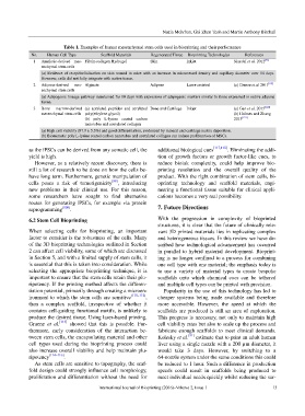
Table 1. Examples of human mesenchymal stem cells used in bioprinting and their performance
No. Human Cell Type Scaffold Materials Regenerated Tissue Bioprinting Technologies References
1 Amniotic-derived mes- Fibrin-collagen Hydrogel Skin Inkjet Skardal et al. 2012 [21]
enchymal stem cells
(a) Evidence of re-epithelialisation on skin wound in mice with an increase in microvessel density and capillary diameter over 14 days.
However, cells did not fully integrate with native tissue.
2 Adipose-derived mes- Alginate Adipose Laser-assisted (a) Gruene et al. 2011 [119]
enchymal stem cells
(a) Adipogenic lineage pathway maintained for 10 days with expression of adipogenic markers similar to those expressed in native adipose
tissue.
3 Bone marrow-derived (a) acrylated peptides and acrylated Bone and Cartilage Inkjet (a) Gao et al. 2015 [120]
mesenchymal stem cells poly(ethylene glycol) (b) Holmes and Zhang
(b) poly L-lysine coated carbon 2013 [121]
nanotubes and acetylated collagen
(a) High cell viability (87.9 ± 5.3%) and good differentiation, evidenced by mineral and cartilage matrix deposition.
(b) Biomimetic poly L-lysine coated carbon nanotubes and acetylated collagen can induce proliferation of MSCs.
as the iPSCs can be derived from any somatic cell, the additional biological cues [117,118] . Eliminating the addi-
yield is high. tion of growth factors or growth factor-like cues, to
However, as a relatively recent discovery, there is reduce bioink complexity, could help improve bio-
still a lot of research to be done on how the cells be- printing resolution and the overall quality of the
have long term. Furthermore, genetic manipulation of product. With the right combination of stem cells, bi-
cells poses a risk of tumorigenicity [98] , introducing oprinting technology and scaffold materials, engi-
new problems in their clinical use. For this reason, neering a functional tissue suitable for clinical appli-
some researchers have sought to find alternative cations becomes a very real possibility.
routes for generating iPSCs, for example via protein
reprogramming [109] . 7. Future Directions
6.2 Stem Cell Bioprinting With the progression in complexity of bioprinted
structures, it is clear that the future of clinically rele-
When selecting cells for bioprinting, an important vant 3D printed materials lies in replicating complex
factor to consider is the robustness of the cells. Many and heterogeneous tissues. In this review we have de-
of the 3D bioprinting technologies outlined in Section scribed how technological advancement has occurred
2 can affect cell viability, some of which are discussed in parallel to hybrid material development. Bioprint-
in Section 5, and with a limited supply of stem cells, it ing is no longer confined to a process for combining
is essential that this is taken into consideration. While one cell type with one material; the emphasis today is
selecting the appropriate bioprinting technique, it is to use a variety of material types to create bespoke
important to ensure that the stem cells retain their plu- scaffolds onto which chemical cues can be tethered
ripotency. If the printing method affects the differen- and multiple cell types can be printed with precision.
tiation potential, primarily through creating a microen- Popularity in the use of this technology has led to
ironment to which the stem cells are sensitive [110–112] , cheaper systems being made available and therefore
then a complex scaffold, irrespective of whether it more accessible. However, the speed at which the
contains cell-guiding functional motifs, is unlikely to scaffolds are produced is still an area of exploration.
produce the desired tissue. Using laser-based printing, This progress is necessary, not only to maintain high
Gruene et al. [113] showed that this is possible. Fur- cell viability rates but also to scale up the process and
thermore, early consideration of the interaction be- fabricate enough scaffolds to meet clinical demands.
tween stem cells, the encapsulating material and other Kolesky et al. [91] estimate that to print an adult human
cell types used during the bioprinting process could liver using a single nozzle with a 200 µm diameter, it
also increase overall viability and help maintain plu- would take 3 days. However, by switching to a
ripotency [114–116] . 64-nozzle system under the same conditions this could
As stem cells are sensitive to topography, the scaf- be reduced to 1 hour. Such a difference in production
fold design could strongly influence cell morphology, speeds could result in scaffolds being produced to
proliferation and differentiation without the need for meet individual needs quickly whilst reducing the sur-
International Journal of Bioprinting (2016)–Volume 2, Issue 1 13