
Page 128 - IJB-10-2
P. 128
International Journal of Bioprinting 3D bioprinting for corneal regeneration
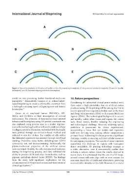
Figure 4. Types of keratoplasty. (A) Structure of healthy cornea, (B) penetrating keratoplasty, (C) deep anterior lamellar keratoplasty, (D) anterior lamellar
keratoplasty, and (E) Descemet stripping endothelial keratoplasty.
model are very promising, further functional studies are 10. Future perspectives
imperative. Alternatively, Campos et al. utilized inkjet-
57
based bioprinting to create a stroma-like construct from Considering the substantial clinical unmet medical need,
a hydrogel containing type I collagen/agarose and human there exists a high probability that an artificial cornea
keratocytes. 93 produced using 3D bioprinting will be among the first to
receive approval from regulatory bodies such as the Food
Goran et al. employed human BM-MSCs, AD- and Drug Administration (FDA) or European Medicines
MSCs, and CS-MSCs in their investigation of corneal Agency (EMA). The technological background is secure,
replacement. The potential of femtosecond laser-assisted and notably, unlike other tissues and organs, the cornea
intrastromal keratoplasty using 3D-printed constructs was lacks blood vessels, thereby reducing the engineering
also explored, using porcine eyes as a model. Alginate- and technological challenge. However, achieving optical
nanocellulose hydrogel, with or without the addition of type perfection is paramount for vision improvement,
1 collagen, served as the matrix. Individual MSC hydrogels necessitating a tissue that can sustain and regenerate
were printed through an extrusion-based method and itself over the long term, making cellular components a
cultured in vitro for 14 days. The viability of cells within primary focus. Addressing this focus presents a significant
the fabricated constructs was assessed through Live/Dead challenge, notably in ensuring sufficient cellular resources
staining, PrestoBlue assay, lactate dehydrogenase (LDH) for autologous procedures. The scarcity of donor numbers
cytotoxicity test, and immunostaining. Additionally, the exacerbates this challenge. In regions with inadequate
physio-mechanical properties of the artificial cornea donor availability, 3D printing technology emerges as
were examined. Notably, the cells demonstrated resilience a viable alternative, even if economically costlier than
during the bioprinting process, and they exhibited the utilizing a cadaver cornea. Given the trends observed in
ability to produce ECM and other biomolecules, such as recent years, it is anticipated that the cost of a 3D tissue-
pigment epithelium-derived factor (PEDF). The findings printed cornea will soon align with or even surpass the
from the study hold significant implications for the economic feasibility of traditional alternatives, providing
advancement of 3D-bioprinted corneas and their potential a considerable stimulus to this field. However, it is crucial
clinical applications. 94
Volume 10 Issue 2 (2024) 120 doi: 10.36922/ijb.1669