
Page 141 - IJB-10-2
P. 141
International Journal of Bioprinting dECM bioink for in vitro disease modeling
assisted bioprinting enables high control precision and realm of 3D models bioprinted with dECM, with special
is not constrained by viscosity or clogging issues, as it emphasis on their medical applications, such as disease
is a nozzle-free method; however, the cellular viability mechanism study and drug testing. In addition, we discuss
is unstable because of heat generation. In summary, it the limitations of the 3D models bioprinted with dECM
27
is necessary to select the suitable bioprinting methods for medical use, along with the relevant improvement
according to designed tissue models. strategies (Figure 1).
Owing to the technical advantages of dECM bioink and
3D bioprinting, dECM-incorporated 3D bioprinting has 2. Decellularized extracellular matrix: key
contributed to the fabrication of precise in vitro models, macromolecules for recapitulating
marked by the recapitulation of microphysiological tissue-specific microenvironment
features of native tissues. Since the development of
3D-bioprinted tissue analogs with dECM bioink by Pati et Decellularized extracellular matrix plays the fundamental
28
al., tissue models bioprinted with dECM have emerged role of preserving the original protein composition
Therefore, the macromolecular
of its native tissue.
14,15
as platforms for drug testing owing to their interactions composition of the dECM and strategies for preserving
with drugs and disease factors, as well as the recapitulation ECM proteins have to be elucidated. In this review, we focus
of tissue-specific pathophysiology. However, despite the
prospects of harmonizing dECM with 3D bioprinting, on ECM proteins rather than carbohydrates or adipose
it is necessary to further investigate dECM bioinks for tissue because of the primary function of ECM proteins
building reliable in vitro models. The bioprinting process as structural supports. In this section, we introduce the
has been significantly improved to build precise structures composition of ECM proteins with respect to the original
and adopt various materials, whereas dECM bioink has tissues, the decellularization methods for the tissues, and
many uncertain features that require investigations, such the function of dECM in tissue engineering.
as protein composition, quality control, safety issues, and 2.1. Composition of decellularized
mechanical properties, which limit the improvement of in extracellular matrix
vitro models and reliability under native tissue condition. The ECMs have different mechanical and biological
Thus, this review focuses on the unmet needs of dECM characteristics depending on their protein compositions
bioinks from the viewpoint of enhancing the utility of and types. They can be roughly categorized into two types
in vitro models for drug testing by incorporating 3D of macromolecules: fibrous proteins, including collagens
bioprinting. First, we describe the components, functions, and elastin; and glycoproteins, including proteoglycans and
29
and fabrication methods of dECMs to elucidate their laminin. The tissue-specific microstructure and biological
physiology. We then discuss the use of 3D bioprinting functions of an ECM are manifested by a combination
technology and dECM, along with their applicability to in of fibrous proteins, glycoproteins, and ECM-associated
vitro modeling. Finally, we address the state of the art in the proteins. A dECM preserves the ECM composition, which
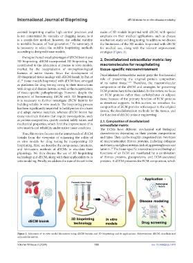
Figure 1. Schematic of in vitro model fabrication using dECM bioinks and 3D bioprinting and its applications. Abbreviations: dECM, decellularized
extracellular matrix.
Volume 10 Issue 2 (2024) 133 doi: 10.36922/ijb.1970