
Page 227 - IJB-10-2
P. 227
International Journal of Bioprinting Property of scaffolds with different lattices
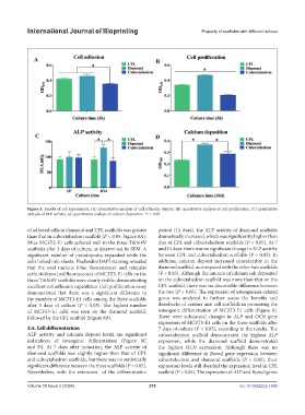
Figure 8. Results of cell experiments. (A) Quantitative analysis of cell adhesion density; (B) quantitative analysis of cell proliferation; (C) quantitative
analysis of ALP activity; (d) quantitative analysis of calcium deposition. *P < 0.05.
of adhered cells in diamond and CPL scaffolds was greater period (14 days), the ALP activity of diamond scaffolds
than that in cuboctahedron scaffold (P < 0.05, Figure 8A). dramatically increased, which was significantly higher than
Mice MC3T3-E1 cells adhered well to the three Ti6Al4V that of CPL and cuboctahedron scaffolds (P < 0.05). At 7
scaffolds after 3 days of culture, as determined by SEM. A and 14 days, there was no significant change in ALP activity
significant number of pseudopodia expanded while the between CPL and cuboctahedron scaffolds (P > 0.05). In
cells linked into sheets. Phalloidin/DAPI staining revealed addition, calcium deposit increased considerably in the
that the oval nucleus (blue fluorescence) and reticular diamond scaffold, as compared with the other two scaffolds
actin skeleton (red fluorescence) of MC3T3-E1 cells on the (P < 0.05). Although the amount of calcium salt deposited
three Ti6Al4V scaffolds were clearly visible, demonstrating on the cuboctahedron scaffold was more than that on the
excellent cell adhesion capabilities. Cell proliferation assay CPL scaffold, there was no discernible difference between
demonstrated that there was a significant difference in the two (P > 0.05). The expression of osteogenesis-related
the number of MC3T3-E1 cells among the three scaffolds genes was analyzed to further assess the benefits and
after 3 days of culture (P < 0.05). The highest number drawbacks of various unit cell scaffolds in promoting the
of MC3T3-E1 cells was seen on the diamond scaffold, osteogenic differentiation of MC3T3-E1 cells (Figure 9).
followed by the CPL scaffold (Figure 8B). There were substantial changes in ALP and OCN gene
expression of MC3T3-E1 cells on the three scaffolds after
3.5. Cell differentiation 7 days of culture (P < 0.05), according to the results. The
ALP activity and calcium deposit levels are significant cuboctahedron scaffold demonstrated the highest ALP
indications of osteogenic differentiation (Figure 8C expression, while the diamond scaffold demonstrated
and D). At 7 days after induction, the ALP activity of the highest OCN expression. Although there was no
diamond scaffolds was slightly higher than that of CPL significant difference in Runx2 gene expression between
and cuboctahedron scaffolds, but there was no statistically cuboctahedron and diamond scaffolds (P > 0.05), their
significant difference between the three scaffolds (P > 0.05). expression levels still dwarfed the expression level in CPL
Nevertheless, with the extension of the differentiation scaffold (P < 0.05). The expression of ALP and Runx2 genes
Volume 10 Issue 2 (2024) 219 doi: 10.36922/ijb.1698