
Page 229 - IJB-10-2
P. 229
International Journal of Bioprinting Property of scaffolds with different lattices
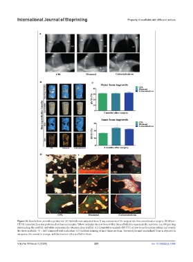
Figure 10. Results from animals experiments. (A) Animals were subjected to an X-ray evaluation of the surgical site three months after surgery. (B) Micro-
CT 3D reconstruction was performed on femoral samples. Yellow indicates the new bone within the scaffold; blue represents the new bone in a 500 µm ring
surrounding the scaffold; and white represents the titanium alloy scaffold. (C) Quantitative analysis (BV/TV) of new bone formation within and outside
the three scaffolds. *P < 0.05 compared with each other. (D) Goldner staining of hard tissue sections. The newly formed mineralized bone is depicted in
turquoise, the osteoid in orange, and the titanium alloy scaffold in black.
Volume 10 Issue 2 (2024) 221 doi: 10.36922/ijb.1698