
Page 363 - IJB-10-2
P. 363
International Journal of Bioprinting 3D bioprinting for vascular regeneration
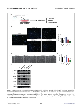
Figure 2. Rapamycin-nanoparticles inhibit smooth muscle cell proliferation and migration. (A) Schematic illustration of the in vitro experimental process.
(B) EdU cell proliferation assay. Scale bars = 500 μm. (C) Quantification of EdU-positive cells. ***P < 0.001; n.s. (not significant), P > 0.05 versus control.
The values represent the mean ± standard deviation (SD) (n = 5). (D) The migration capacity of human coronary artery smooth muscle cells treated with
rapamycin and rapamycin-nanoparticles is investigated using a scratched wound healing assay. (E) Quantification of migrated area. *P < 0.05; ***P <
0.001; n.s., P > 0.05 versus control. The values represent mean ± SD (n = 5). (F) Protein expression of p-mTOR, mTOR, p-S6K1, S6K1, p-AKT, and AKT
as evaluated by Western blotting. Abbreviation: NP, nanoparticle.
Volume 10 Issue 2 (2024) 355 doi: 10.36922/ijb.1465