
Page 365 - IJB-10-2
P. 365
International Journal of Bioprinting 3D bioprinting for vascular regeneration
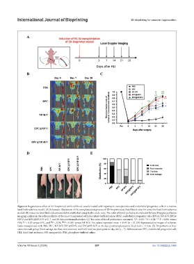
Figure 4. Regenerative effect of 3D-bioprinted artificial blood vessels loaded with rapamycin-nanoparticles and endothelial progenitor cells in a murine
hind limb ischemia model. (A) Schematic illustration of the transplantation process of 3D-bioprinted artificial blood vessel in a murine hind limb ischemia
model. (B) A murine hind limb ischemia model is established using Balb/c nude mice. The ratio of blood perfusion is evaluated by laser Doppler perfusion
imaging analysis in the ischemic limbs of the mice transplanted with phosphate-buffered saline (PBS), endothelial progenitor cells (EPCs), NP-R/V, EPC@
NP/V, and EPC@NP-R/V at 0, 7, and 28 days posttransplantation. (C) The ratio of blood perfusion is measured. *P < 0.05; **P < 0.01; ***P < 0.001 versus
#
$$
$$$
PBS; P < 0.05 versus EPC, and P < 0.01; P < 0.001 versus NP-R/V. The values represent mean ± SEM (n = 5). (D) Representative images of ischemic
limbs transplanted with PBS, EPC, NP-R/V, EPC@NP/V, and EPC@NP-R/V at 28 days posttransplantation. Scale bars = 5 mm. (E) Proportions of four
states for each group (limb salvage, toe loss, foot necrosis, and limb loss) on postoperative day 28 (n = 7). Abbreviations: EPC, endothelial progenitor cell;
HLI, hind limb ischemia; NP, nanoparticle; PBS, phosphate-buffered saline.
Volume 10 Issue 2 (2024) 357 doi: 10.36922/ijb.1465