Page 408 - IJB-10-2
P. 408
International Journal of Bioprinting                            3D-bioprinted macrophage inflammation model


            bioprinting. Different concentrations of GelMA (2.5,   of FDA-bound cells within the total cell population (both
            5, 7.5, 10, 12.5, 15% (w/v)) containing 0.15% (w/v)   FDA and PI-strained cells).
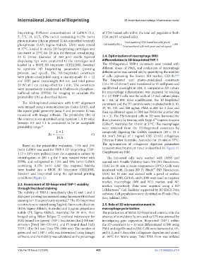
            photoinitiator lithium phenyl (2,4,6-trimethyl benzoyl)         TotalnumberofFDA boundlivecells green(  )
            phosphinate (LAP; Sigma-Aldrich, USA) were mixed    Cellviability(%)   Totall numberof cells with green andred signals  100
            at 37°C, loaded in sterile 3D bioprinting cartridges and
            incubated at 23°C for 20 min for thermal crosslinking.
            25 G (inner diameter of 260 µm) sterile tapered    2.4. Optimization of macrophage (M0)
            dispensing tips were connected to the cartridges and   differentiation in 3D-bioprinted THP-1
            loaded  in  a  BIOX  3D  bioprinter  (CELLINK,  Sweden)   The  3D-bioprinted THP-1  constructs  were treated  with
            to  optimize  3D  bioprinting  parameters  (printing   different doses of PMA, and evaluation of macrophage
            pressure and speed). The 3D-bioprinted constructs   differentiation was carried out by quantifying the number
            were photo-crosslinked using a custom-made 15 × 12   of cells expressing the known M0 marker, CD11b. 24-26
            cm  LED panel (wavelength 405 nm and total power   The bioprinted and photo-crosslinked constructs
              2
                                                                              3
            20 W) at 1 cm curing offset for 1 min. The constructs   (10 × 10 × 0.8 mm ) were transferred to 12-well plates and
            were immediately transferred to Dulbecco’s phosphate-  equilibrated overnight in BM. A comparative 2D culture
            buffered saline (DPBS) for imaging to calculate the   for  macrophage differentiation  was  prepared  by seeding
                                                                    5
            printability (Pr) as described below.              5 × 10  THP-1 cells into the wells of 12-well culture plates
                                                               in 1 mL of BM. After equilibration, the 3D-bioprinted
               The 3D-bioprinted constructs with 0–90° alignment   constructs and the 2D controls were incubated with 0, 10,
            were imaged using a stereomicroscope (Leica EZ4E), and   25, 50, 100, and 200 ng/mL PMA in BM for 2 days and
            the square grids’ geometry (area A and perimeter L) was   then equilibrated again in BM (no PMA) for another day
            measured with ImageJ software. The printability (Pr) of   (n = 3). The PMA-treated cells in 2D were harvested for
            the constructs was quantified using Equation I. A Pr value   flow cytometry by treating with TrypLE  Express Enzyme
                                                                                               ™
            between 0.9 and 1.1  is considered to be  an acceptable   (Gibco , Australia) for 10 min at 37°C. The macrophages
                                                                    ®
            printability range. 22                             were  retrieved from  the  3D-bioprinted constructs  by
                                                               completely digesting the GelMA constructs (10  × 10  ×
                                                        (I)    0.8 mm ) 200 µL of 1 mg/mL (285 U/mL) collagenase
                                                                     3
                                                               (Thermo Fisher Scientific, Australia) for 10 min at 37°C.
               Based  on  the  printability  evaluation,  7.5%  and  10%   The optimization of collagenase digestion parameters
            (w/v) GelMA was used for THP-1 3D bioprinting. THP-  (concentration/treatment time) is described in Figure S1
                   6
            1 (2 × 10 ) were pelleted from the suspension culture by   (Supplementary File).
            centrifugation  at 200 ×  g for  3 min, washed  twice with   The harvested cells were washed with DPBS and
            DPBS,  and redispersed in  7.5%  and  10%  (w/v)  GelMA   stained with Fixable Viability Stain 510 (BD Biosciences,
            containing 0.15% (w/v) LAP. The prepared bioinks   USA) for 20 min at room temperature (23°C). Cells were
            were loaded into a BIOX 3D bioprinter (CELLINK,    incubated with Human BD Fc Block  (BD Biosciences,
                                                                                              ™
            Sweden) and bioprinted using the optimized printing    USA) for 10 min and stained with a panel of surface
            conditions (Figure 1).                             markers. CD90, CD11b, and CD80 were used as negative
                                                               marker, macrophage (M0 and M1) marker, and M1
            2.3. Assessment of 3D-bioprinted THP-1 viability   marker, respectively. Data were  acquired using a BD
            through live/dead staining                         LSRFortessa  Cell Analyzer supported by BD FACS Diva
                                                                         ™
            The viability of THP-1 immediately (day 0) and 1 and 3   software. Cell populations were identified on FlowJo (Tree
            days post-printing was assessed using fluorescent live/dead   Star, Ashland, OR).
            staining (n = 3) as previously reported.  The 3D-bioprinted
                                          23
            constructs were stained using 5 µg/mL fluorescein diacetate   2.5. Role of 3D microenvironment in
            (FDA; Sigma-Aldrich, Australia) and 2 μg/mL propidium   macrophage polarization
            iodide  (PI;  Sigma-Aldrich,  Australia)  for  30  min,  then   The polarization of M0 in 3D-bioprinted constructs in the
            imaged using Nikon Eclipse Ti confocal microscope for   absence of stimulatory factors such as LPS was assessed by
            FDA-bound live (green) THP-1 (excitation [Exc] 488 nm/  investigating  gene  expression.  Bioprinted  THP-1  within
            emission [Ems] 500–550 nm), and PI-bound dead (red)   the 3D constructs (n = 3) were differentiated to M0 using
            THP-1 (Exc 561 nm / Ems 570–1000 nm). The number of   PMA and equilibrated in BM. Cells were harvested at 6 h,
            green and red THP-1 cells was determined using ImageJ   and 1, 2, and 4 days after collagenase digestion and stored
            software, and the viability was calculated as the percentage   at -80°C for future assay. Total RNA from was isolated


            Volume 10 Issue 2 (2024)                       400                                doi: 10.36922/ijb.2116

