
Page 409 - IJB-10-2
P. 409
International Journal of Bioprinting 3D-bioprinted macrophage inflammation model
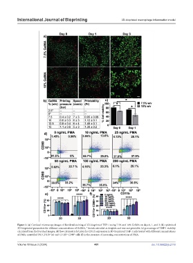
Figure 1. (a) Confocal microscopy images of live/dead staining of 3D-bioprinted THP-1 using 7.5% and 10% GelMA on days 0, 1, and 3; (b) optimized
3D-bioprinted parameters for different concentrations of GelMA; * bioink extruded as droplets and was not printable; (c) percentage of THP-1 viability
calculated from the live/dead images; (d) flow cytometry dot plots for CD11b expression in 3D-bioprinted THP-1 cells treated with different concentrations
of PMA; quantified (%) CD11b (e) and CD11b CD80 cells (f) in the presence of increasing concentrations of PMA.
+
+
+
Volume 10 Issue 2 (2024) 401 doi: 10.36922/ijb.2116