
Page 415 - IJB-10-2
P. 415
International Journal of Bioprinting 3D-bioprinted macrophage inflammation model
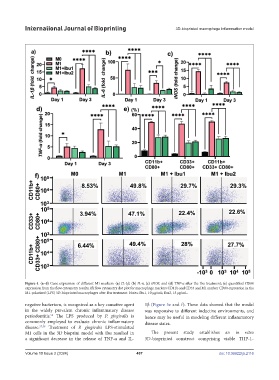
Figure 4. (a–d) Gene expression of different M1 markers: (a) IL-1β, (b) IL-6, (c) iNOS, and (d) TNF-α after the Ibu treatment; (e) quantified CD80
expression from the flow cytometry results; (f) flow cytometry dot plot for macrophage markers CD11b and CD33 and M1 marker CD80 expression in the
M1-polarized (LPS) 3D-bioprinted macrophages after Ibu treatment. Notes: Ibu1, 10 µg/mL; Ibu2, 15 µg/mL.
negative bacterium, is recognized as a key causative agent 1β (Figure 5e and f). These data showed that the model
in the widely prevalent chronic inflammatory disease was responsive to different inductive environments, and
periodontitis. The LPS produced by P. gingivalis is hence may be useful in modeling different inflammatory
56
commonly employed to evaluate chronic inflammatory disease states.
disease. 57,58 Treatment of P. gingivalis LPS-stimulated
M1 cells in the 3D bioprint model with Ibu resulted in The present study establishes an in vitro
a significant decrease in the release of TNF-α and IL- 3D-bioprinted construct comprising viable THP-1-
Volume 10 Issue 2 (2024) 407 doi: 10.36922/ijb.2116