
Page 11 - IJB-3-1
P. 11
Andy Wen Loong Liew and Yilei Zhang
ciples but slightly different methodology, was carried removal process. Many reports found in literature to-
out where researchers were able to incorporate an in- day utilize the same fugitive ink approach to tackle the
terconnected vascular network within bulk hydrogel problem of vascularization [39] (Figure 2). Fugitive ink
containing hepatocytes and showed that perfusion of may contain cytotoxic compounds which are detri-
the vascular network with cell medium was able to mental to cell viability and affect cell phenotype. The
sustain metabolic activity of the surrounding hepato- process of removing the fugitive ink, such as chemical
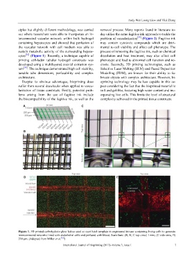
cytes [32] (Figure 1). Recently, a technique capable of dissolution and heat treatment, may also affect cell
printing cell-laden tubular hydrogel constructs was phenotype and lead to abnormal cell function and ne-
developed using a multilayered coaxial extrusion sys- crosis. Secondly, 3D printing technologies, such as
tem [38] . The technique demonstrated high cell viability, Selective Laser Melting (SLM) and Fused Deposition
tunable tube dimensions, perfusability and complex Modelling (FDM), are known for their ability to fa-
architecture. bricate objects with complex architecture. However, bi-
Despite its obvious advantages, bioprinting does oprinting technology may be less capable in this as-
suffer from several drawbacks when applied to vascu- pect considering the fact that the bioprinted material is
larization of tissue constructs. Firstly, potential prob- soft and gel-like, featuring high water content and inc-
lems arising from the use of fugitive ink include orporating live cells. This limits the level of structural
the biocompatibility of the fugitive ink, as well as the complexity achieved in the printed tissue constructs.
Figure 1. 3D printed carbohydrate-glass lattice used as sacrificial template in engineered tissues containing living cells to generate
interconnected networks lined with endothelial cells and perfused with blood. Scale bars: (B, D, C top-view) 1 mm; (C side-view, E)
200 μm. (Adopted from Miller et al. [32] )
International Journal of Bioprinting (2017)–Volume 3, Issue 1 7