
Page 88 - IJB-3-1
P. 88
Roles of support materials in 3D bioprinting — Present and future
for bioprinted 3D complex hollow structures. ing a human organ. This is due to the fact that only
one of the materials, either model or support materials
2. Roles of Support Materials in Bioprinting (mostly support material), has the good mechanical
Recently, printing beyond 2.5D (2.5D shape is the strength which is responsible for the shape stability
shape that comes from repeatedly printing a 2D pat- and shape integrity.
tern in Z direction without changing the pattern in any Another recent advancement is the development of
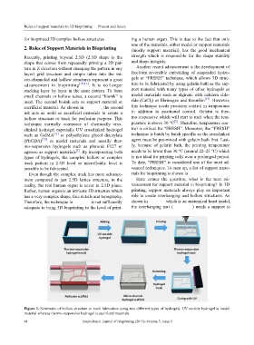
layer) grid structure and simple tubes into the mi- freeform reversible embedding of suspended hydro-
cro-channeled and hollow structures represent a great gels or “FRESH” technique, which allows 3D struc-
advancement in bioprinting [10,11] . It is no l onger ture to be fabricated by using gelatin bath as the sup-
stacking layer by layer in the same pattern. To form port material with many types of other hydrogels as
small channels or hollow tubes, a second “bioink” is model materials such as alginate with calcium chlo-
[13]
used. The second bioink acts as support material or ride (CaCl 2) or fibrinogen and thrombin . However,
sacrificial material. As shown in Figure 1, the second this technique needs precision control in temperature
ink acts as mold or sacrificial materials to create a in addition to positional control. Gelatin is ther-
hollow structure or track for perfusion purpose. This mo-responsive which will start to melt when the tem-
[2]
technique normally composes of chemically cros- perature is above 30 °C . Therefore, temperature con-
slinked hydrogel especially UV crosslinked hydrogel trol is critical for “FRESH”. Moreover, the “FRESH”
such as GelMA [11] or polyethylene glycol diacrylate technique is batch by batch specific so the crosslinked
(PEGDA) [12] as model materials and usually ther- agent must be pre-mixed with gelatin bath first. Last-
mo-responsive hydrogels such as pluronic F127 or ly, because of gelatin bath, the printing temperature
[2]
agarose as support materials . By incorporating both needs to be lower than 30 °C (around 22–25 °C) which
types of hydrogels, the complex hollow or complex is not ideal for printing cells over a prolonged period.
track pattern in 2.5D level or microfluidic level is To date, “FRESH” is considered one of the most ad-
possible to be fabricated. vanced techniques. To sum up, a list of support mate-
Even though the complex track has more advance- rials for bioprinting is shown in Table 1.
ment compared to just 2.5D lattice structure, in the Here comes the question, what is the next ad-
reality, the real human organ is never in 2.5D plane. vancement for support material in bioprinting? In 3D
Rather, human organ is an intricate 3D structure which printing, support materials always play an important
has a very complex shape, fine details and topography. role to create overhanging and hollow structures. As
Therefore, the technique in Figure 1 is not sufficiently shown in Figure 2 which is an anatomical heart model,
adequate to bring 3D bioprinting to the level of print- the overhanging part (Figure 2A) needs a support to
Figure 1. Schematic of hollow structure or track fabrication using two different types of hydrogels. UV curable hydrogel is model
material whereas thermo-responsive hydrogel is sacrificial materials.
84 International Journal of Bioprinting (2017)–Volume 3, Issue 1