
Page 67 - IJB-3-2
P. 67
Roger Sachan, et. al.
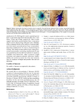
Figure 6. Optical micrographs showing the insertion sites in surgically discarded human abdominal skin for (a) an uncoated polyglycolic
acid microneedle array, (b) a matrix-assisted pulsed laser evaporation-coated microneedle array from deposition with the AmfB(260)
target (amphotericin B 1040 mg/mL + 1% polyvinylpyrrolidone), and (c) a matrix-assisted pulsed laser evaporation-coated microneedle
array from deposition with the AmfB(520) target (amphotericin B 2080 mg/mL + 1% polyvinylpyrrolidone). The location of microneedle
insertion was identified using methylene blue dye.
amphotericin B (2080 mg/mL) matrix-assisted pulsed 2. Torrado J J, Espada R, Ballesteros M P, et al., 2008, Ampho-
laser evaporation-deposited microneedle array were tericin B formulations and drug targeting. Journal of Pharma
0 mm, 11 mm, and 18 mm, respectively. These results ceutical Sciences, vol.97(7): 2405–2425.
sug gest that matrix-assisted pulsed laser evaporation
may be used to deposit drugs with poor water solubility, https://dx.doi.org/10.1002/jps.21179
such as amphotericin B on the surfaces of microneedles, 3. Trejo W H and Bennett R E, 1963, Streptomyces nodosus
and that matrix-assisted pulsed laser evaporation- sp. nov., the amphotericin-producing organism. Journal of
deposited amphotericin B retains pharmacological Bacteriology, vol.85(2): 436–439.
activity. The matrix-assisted pulsed laser evaporation- 4. Hamill R J, 2013, Amphotericin B formulations: A compa-
coated microneedles containing amphotericin B may
have potential use for transdermal treatment of cutaneous rative review of efficacy and toxicity. Drugs, vol.73(9): 919–
Candida yeast infections, other cutaneous fungal 934.
infections, and cutaneous parasitic infections. Further https://dx.doi.org/10.1007/s40265-013-0069-4
studies are needed to assess the dose and exposure 5. Laniado-Laborin R and Cabrales-Vargas M N, 2009, Ampho-
time for treatment of fungal and parasitic skin and nail tericin B: Side effects and toxicity. Revista Iberoa mericana de
infections.
Micología, vol.26(4): 223–227.
Conflict of Interest https://dx.doi.org/10.1016/j.riam.2009.06.003
No conflict of interest was reported by the authors. 6. Khanna P, Strom J A, Malone J I, et al., 2008, Microneedle-
Acknowledgments based automated therapy for diabetes mellitus. Journal of
Diabetes Science and Technology, vol.2(6): 1122–1129.
We would like to acknowledge C Mooney (NCSU https:/dx./doi.org/10.1177/193229680800200621
Analytical Instrumentation Facility) for his aid with 7. Baria S H, Gohel M C, Mehta T A, et al., 2011, Microneedles:
electron microscopy, B Andersen for her aid with Fourier
transform infared spectrosocpy (NCSU College of An emerging transdermal drug delivery system. Journal of
Textiles), P Strader (NCSU Analytical Instrumentation Pharmacology and Pharmacotherapeutics, vol.64(1): 11–29.
Facility) for his aid with nanoindentation, the US https://dx.doi.org/10.1111/j.2042-7158.2011.01369.x
National Institutes of Health (Award # 1R21AI117748- 8. Arora A, Prausnitz M R, Mitragotri S, 2008, Micro-scale
01A1), the US National Science Foundation (Award devices for transdermal drug delivery. International Journal
CMMI 1258536), and the US Office of Naval Research
(Award # N00014-15-1-2323). of Pharmaceutics, vol.364(2): 227–236.
https://dx.doi.org/10.1016/j.ijpharm.2008.08.032
References 9. Gill H S, Denson D D, Burris B A, et al., 2008, Effect of
1. Ostrosky-Zeichner L, Marr K A, Rex J H, et al., 2003, Am- microneedle design on pain in human volunteers. The Clinical
pho tericin B: Time for a new “gold standard”. Clinical Journal of Pain, vol.24(7): 585–594.
Infectious Diseases, vol.37(3): 415–425. https://dx.doi.org/10.1097/AJP.0b013e31816778f9
https://dx.doi.org/10.1086/376634 10. Nahar M, Mishra D, Dubey V, et al., 2008, Development,
International Journal of Bioprinting (2017)–Volume 3, Issue 2 155