
Page 63 - IJB-3-2
P. 63
Roger Sachan, et. al.
buffered saline (10×) (VWR International, West Chester, elastic modulus values for the uncoated polyglycolic
PA, USA). Overnight broth cultures of Candida albicans acid material (Table 2). Taking into account the
with yeast nitrogen base and 100 mM dextrose were Poisson’s ratio of the diamond indenter tip (0.07) and
prepared. Cell pellets were obtained using centrifugation assum ing a Poisson’s ratio for the polyglycolic acid
(4500 rpm) for 10 min; these pellets were subsequently material of 0.3, the nanoindentation study indicated that
8
resuspended to a cell density of approximately 10 cells/ the polyglycolic acid material had a reduced Young’s
ml in phosphate-buffered saline (PBS) (1×); PBS (10×) modulus value of approximately 5.5 GPa and a hardness
was diluted using deionized water to create PBS (1×). value of approximately 230 MPa. Park et al. evaluated
Agar plates were inoculated with Candida albicans the mechanical parameters of microneedle materials
cultures following resuspension of the cell pellets. and suggested that microneedle materials with Young’s
Sabouraud dextrose agar was swabbed with Candida modulus values higher than ~1 GPa were associated with
albicans. Triphenyltetrazolium chloride was added fracture forces that surpassed skin insertion forces [35] .
into each agar plate to serve as a visualization aid; The nanoindentation results indicate that the polyglycolic
this dye turns red in color in the presence of microbial acid material has sufficient stiffness to penetrate the skin.
growth [29–33] . The plates were incubated at 37 °C for 24 Figure 2 shows the Fourier transform infrared spectra
hours. After 24 hours, the plates were evaluated for of matrix-assisted pulsed laser evaporation-deposited
regions of inhibited microbial growth. coatings on glass. Figure 2(a) shows the spectrum for
deposition with the AmfB(260) target (amphotericin B
2.9 Skin Penetration Properties of the Micro- 1040 mg/mL + 1% polyvinylpyrrolidone) and Figure
needle Arrays 2(b) shows the spectrum for deposition with the
Discarded human abdominal skin is commonly used to AmfB(520) target (amphotericin B 2080 mg/mL + 1%
assess the skin penetration properties of microneedle polyvinylpyrrolidone). The contribution of amphotericin
arrays [34] . Methylene blue was used to examine the B to the spectra is associated with N–H (overlapped
−1
−1
peak around 670 cm ), C–H (around 750 cm ), C–O
pores in the human abdominal skin that were created stretching (around 1,380 cm ), C=C stretching (around
−1
by the microneedle arrays. Surgically discarded human −1 −1
abdominal skin was obtained from Duke Hospital, USA, 1600 cm ), C–H stretching (around 3000 cm ), and O–
−1 [36]
in accordance with an institutionally approved IRB H stretching (around 3350 cm ) . The contribution of
polyvinylpyrrolidone to the spectra is associated with a
protocol (DNOR 80 1185-01); the skin was processed strong band around 1660 cm ; this band is assigned to
−1
with Zimmer Air Dermatome prior to use. The split- the amide carbonyl group of N-vinyl-2-pyrrolidone [37] .
thickness skin pieces was preloaded with 200 µL of Other bands associated with polyvinylpyrrolidone in the
1% methylene blue dissolved in water and punched spectra are around 1380 cm , which is assigned to bond
−1
with the uncoated polyglycolic acid microneedle, vibrations of the NO group, and around 1290 cm ,
−
−1
3
the matrix-assisted pulsed laser evaporation-coated which is assigned to N–OH bond vibrations . A major
[29]
microneedle from deposition with the AmfB(260) absorption band is located at around 1050 cm , which
−1
target, or the matrix-assisted pulsed laser evaporation- is attributed to dimethyl sulfoxide’s S–O stretching [37] .
coated microneedle from deposition with the AmfB(520) The results indicate that the chemical functionality of
target. The microneedle assembly was held using a the matrix-assisted pulsed laser evaporation-deposited
hemostatic forceps to help control the penetration of coatings is similar to those of the starting materials. The
the microneedles into the skin. Bright field images were spectral features for the matrix-assisted pulsed laser
obtained with the Olympus imaging system around evaporation-deposited coating do not show a noticeable
30 min to 1 h after the punch procedure. departure (indicative of chemical modification) from the
3. Results and Discussion starting materials.
Figure 3 shows scanning electron microscopy
Nanoindentation was used to obtain the hardness and images of unmodified and matrix-assisted pulsed laser
evaporation-modified polyglycolic acid microneedles.
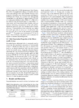
Table 2. Nanoindentation result obtained from the base of an
uncoated polyglycolic acid microneedle. Reduced modulus (E r ) Figure 3(a), (b), and (c) show scanning electron
and hardness (H) data were obtained from nanoindentation data micrographs of an uncoated polyglycolic acid micro-
using Oliver-Pharr analysis. needle, a scanning electron micrograph of a matrix-
Data Indent 1 Indent 2 assisted pulsed laser evaporation-coated microneedle
Reduced modulus (E r ) 5.61 GPa 5.43 GPa from deposition with the AmfB(260) target (amphotericin
B 1040 mg/mL + 1% polyvinylpyrrolidone), and a
Hardness (H) 238.96 MPa 217.37 MPa
scanning electron micrograph of a matrix-assisted
Maximum depth 287.5 nm 299.1 nm pulsed laser evaporation-coated microneedle from
International Journal of Bioprinting (2017)–Volume 3, Issue 2 151