
Page 62 - IJB-3-2
P. 62
Printing amphotericin B on microneedles using matrix-assisted pulsed laser evaporation
the matrix-assisted pulsed laser evaporation procedure 2.6 3D Laser Scanning Confocal Microscopy of
by exposure to ultraviolet light from a VL-115 UV lamp. the Microneedle Arrays
The parameters used for matrix-assisted pulsed laser
evaporation of amphotericin B on the surfaces of the The surfaces of the unmodified and matrix-assisted
silicon <100> wafer substrates, optic glass substrates, pulsed laser evaporation-coated microneedles were
and polyglycolic acid microneedles are shown in Table evaluated using a VK-X250 3D laser scanning confocal
1. All of the matrix-assisted pulsed laser evaporation microscope (Keyence, Tokyo, Japan). In this instrument,
depositions were conducted with a KrF* excimer laser the laser was rastered in an XY pattern across the
source using a wavelength of 248 nm, a repetition rate of field of view, and 0.5 nm steps in the Z-direction were
10 Hz, a pulse duration of 25 ns, and an optimum laser obtained. The 16-bit photomultiplier receiving element
2
fluence of 300 mJ/cm ; these parameters are commonly was dynamically latched onto the highest reflected laser
used for matrix-assisted pulsed laser evaporation of intensity for each pixel. At that point, it set a color value
and height value to create a three-dimensional fully-
coatings [26−28] . 150,000 laser pulses were used for each in-focus topo graphical map. This laser-based approach
deposition. Both the substrate and the target were rotated enables data acquisition from complex surface shapes.
at a rate of 50 Hz during the depositions. The laser beam
scanned the entire surface of the target at an angle of 2.7 Fourier Transform Infrared Spectra of
45°. During the matrix-assisted pulsed laser evaporation Matrix-Assisted Pulsed Laser Evaporation-
process, the rotating target was maintained in direct Depo sited Coatings on Glass
contact with a cooling apparatus, which included a liquid
nitrogen reservoir and was connected to the target with The materials that were processed using matrix-
copper pipes. The target was maintained at a temperature assisted pulsed laser evaporation were examined with
of ~173 K using active liquid nitrogen cooling. Using Fourier transform infrared spectroscopy to determine
this setup, rapid evaporation of matrix-assisted pulsed if the functional groups of the matrix-assisted pulsed
laser evaporation target inside the deposition chamber laser evaporation target materials were identifiable in
is significantly decreased. All of the depositions were the matrix-assisted pulsed laser evaporation-coated
−1
performed using a background pressure of 2 × 10 Pa surfaces. The Fourier transform infrared spectra were
and a substrate-to-target separation distance of 5 cm. obtained using a Nexus 470 system, which included an
™
OMNI sampler, a continuum microscope, and OMNIC
A laser beam homogenizer was used for improving the analysis software (Thermo Fisher, Waltham, MA, USA).
energy distribution of the laser spot and for increasing
the coated region on the substrate. 2.8 Modified Agar Disk Diffusion Assay of the
2.5 Variable Pressure Scanning Electron Microneedle Arrays
Micro scopy of the Microneedle Arrays A modified agar disk diffusion assay was used to
examine the growth-inhibiting effects of (a) the matrix-
An S-3200 variable-pressure scanning electron micro- assisted pulsed laser evaporation-coated microneedle
scope with an energy-dispersive X-ray spec trometer array from deposition with the AmfB(260) target and
(Hitachi, Tokyo, Japan) was used to obtain imaging data (b) the matrix-assisted pulsed laser evaporation-coated
and energy-dispersive X-ray spectra from the unmodified microneedle array from deposition with the AmfB(520)
and matrix-assisted pulsed laser evaporation-coated target; cultures of Candida albicans (ATCC 90028)
microneedle arrays. Prior to imaging, the unmodified (American Type Culture Collection, Manassas, VA,
and matrix-assisted pulsed laser evaporation-coated USA) were used in this study [11,12] . Matrix-assisted
microneedle arrays were sputter-coated with a layer pulsed laser evaporation-coated silicon <100> wafer
of 60% gold–40% palladium for three minutes in a sub strates and optic glass substrates were also evaluated
Technics Hummer II system (Anatech, Battle Creek, in this study. The reagents used for the microbial cultures
MI, USA). The energy-dispersive X-ray spectra were included yeast nitrogen base, Sabouraud dextrose agar,
obtained in charge reduction mode. triphenyltetrazolium chloride, dextrose, and phosphate-
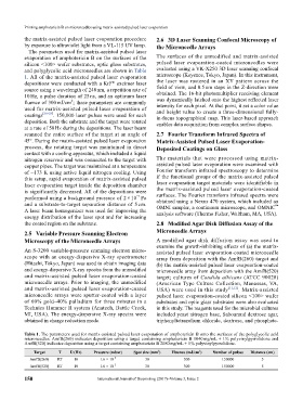
Table 1. The parameters used for matrix-assisted pulsed laser evaporation of amphotericin B onto the surfaces of the polyglycolic acid
microneedles. AmfB(260) indicates deposition using a target containing amphotericin B 1040 mg/mL + 1% polyvinylpyrrolidone and
AmfB(520) indicates deposition using a target containing amphotericin B 2080 mg/mL + 1% polyvinylpyrrolidone.
2
Target T Ʋ (Hz) Pressure (mbar) Spot size (mm ) Fluence (mJ/cm ) Number of pulses Distance (cm)
2
AmfB(260) RT 10 1.6 × 10 −2 30 300 150000 5
AmfB(520) RT 10 1.6 × 10 −2 30 300 150000 5
150 International Journal of Bioprinting (2017)–Volume 3, Issue 2