
Page 241 - IJB-10-3
P. 241
International Journal of Bioprinting Increased ECM stiffness enhances chemoresistance
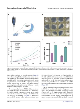
Figure 2. Fabrication of 3D-bioprinted ovarian cancer model. (A) Schematic illustration of the 3D bioprinting process. (B) An image of the 3DP-Ov
model. Scale bar: 200 μm. (C) Representative stress-strain curves of 3D models under different conditions. (D) Young’s modulus of 3D models under
different conditions.
high resolution tailored for research purposes. Figure 2B obviously different. To be specific, the Young’s moduli of
shows the resulting 3D-bioprinted ovarian cancer model. 6%-3DP and 8%-3DP tumor models were 0.009±0.001
Our preliminary study involved exploring suitable bioink MPa and 0.024±0.001 MPa with a significant difference.
materials and 3D bioprinting technologies for cell growth Additionally, for the 3D-bioprinted models without tumor
and proliferation. We used GelMA, a hydrogel containing cells, the Young’s modulus of 6% GelMA and 8% GelMA
23
ovarian cancer cells, to print a 3D lattice structure were 0.015±0.003 MPa and 0.027±0.002 MPa, respectively
measuring 6 mm in length, 6 mm in width, and 1 mm in (Figure 2D).
height, according to a preset procedure. The 3D-bioprinted ovarian cancer model was cultured
To investigate the impact of ECM stiffness on tumor in SKOV3 cell medium with medium changes every two
cells, we mixed tumor cell suspensions with 10% GelMA days. C-AM/PI staining was used to evaluate the viability
hydrogel in suitable proportions to create tumor models of tumor cells in 3D ovarian cancer model. The results
with GelMA concentrations of 6% (i.e., 6%-3DP) and 8% revealed that cell viability of 6%-3DP was approximately
(i.e., 8%-3DP). As presented in Figure 2C, stress-strain 82% on the first day after printing. With the extension of
curves of 3D models under various conditions were culture time after printing, cell viability gradually increased
Volume 10 Issue 3 (2024) 233 doi: 10.36922/ijb.1673