
Page 242 - IJB-10-3
P. 242
International Journal of Bioprinting Increased ECM stiffness enhances chemoresistance
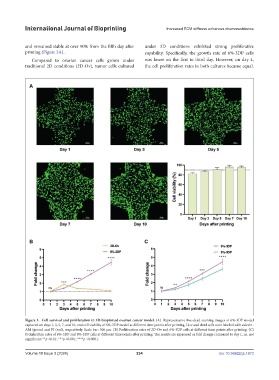
and remained stable at over 90% from the fifth day after under 3D conditions exhibited strong proliferative
printing (Figure 3A). capability. Specifically, the growth rate of 6%-3DP cells
Compared to ovarian cancer cells grown under was lower on the first to third day. However, on day 4,
traditional 2D conditions (2D-Ov), tumor cells cultured the cell proliferation rates in both cultures became equal.
Figure 3. Cell survival and proliferation in 3D-bioprinted ovarian cancer model. (A) Representative live-dead staining images of 6%-3DP model
captured on days 1, 3, 5, 7, and 10, and cell viability of 6%-3DP model at different time points after printing. Live and dead cells were labeled with calcein-
AM (green) and PI (red), respectively. Scale bar: 500 μm. (B) Proliferation rates of 2D-Ov and 6%-3DP cells at different time points after printing. (C)
Proliferation rates of 6%-3DP and 8%-3DP cells at different time points after printing. The results are expressed as fold change compared to day 1. ns, not
significant; **p <0.01; ***p <0.001; ****p <0.0001.
Volume 10 Issue 3 (2024) 234 doi: 10.36922/ijb.1673