
Page 246 - IJB-10-3
P. 246
International Journal of Bioprinting Increased ECM stiffness enhances chemoresistance
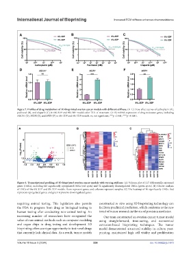
Figure 7. Profiles of drug metabolism of 3D-bioprinted ovarian cancer models with different stiffness. (A–C) Dose-effect curves of carboplatin (A),
paclitaxel (B), and olaparib (C) in 6%-3DP and 8%-3DP models after 72 h of treatment. (D–F) mRNA expression of drug resistance genes, including
ABCA1 (D), MDR1(E), and MRP1(F) in 6%-3DP and 8%-3DP models. ns, not significant; ***p <0.001; ****p <0.0001.
Figure 8. Transcriptional profiling of 3D-bioprinted ovarian cancer models with varying stiffness. (A) Volcano plot of 217 differentially expressed
genes (DEGs), including 145 significantly upregulated DEGs (red spots) and 72 significantly downregulated DEGs (green spots). (B) Cluster analysis
of DEGs of the 6%-3DP and 8%-3DP models. Rows represent genes, and columns represent samples. (C) The heatmap of 16 significantly DEGs. Red
represents upregulated genes, and green represents downregulated genes.
requiring animal testing. This legislation also permits constructed in vitro using 3D bioprinting technology can
the FDA to progress from drug or biological testing to facilitate preclinical prediction, which conforms to the new
human testing after conducting non-animal testing. An trend of tumor research in the era of precision medicine.
increasing number of researchers have recognized the Our team constructed an ovarian cancer tumor model
value of non-animal methods such as computer modeling using straightforward, time-saving, and economical
and organ chips in drug testing and development. 3D extrusion-based bioprinting techniques. The tumor
bioprinting offers a unique opportunity to test novel drugs model demonstrated structural stability in culture post-
that currently lack clinical data. As a result, tumor models printing, maintained high cell vitality and proliferation
Volume 10 Issue 3 (2024) 238 doi: 10.36922/ijb.1673