
Page 255 - IJB-10-3
P. 255
International Journal of Bioprinting Bioprinting organoids for toxicity testing
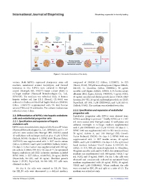
Figure 1. Schematic illustration of the study.
vectors. Both hiPSCs expressed pluripotent stem cell composed of DMEM-F12 (Gibco, 11320033), 1× ITS
markers, maintained normal karyotypes, and formed (Merck, I3146), 500 μM monothioglycerol (Sigma-Aldrich,
teratomas in vivo. hiPSCs were cultured in Matrigel- M6145), 1× GlutaMax (Gibco, 35050061), 50 μg/mL
coated (Matrigel, BD, 354277) tissue culture plates in ascorbic acid (Sigma-Aldrich, A8960), 0.1% bovine serum
ncTarget medium (Nuwacell Biotechnologies Co., Ltd., albumin (BSA; Sigma-Aldrich, V900933), 5 ng/mL FGF2,
RP01020). The medium was refreshed daily. A human 10 ng/mL vascular endothelial growth factor (VEGF; R&D
hepatic stellate cell line LX-2 (Procell, CL-0560) was Systems, 293-VE), 20 ng/mL epidermal growth factor (EGF;
cultured in Dulbecco’s Modified Eagle’s Medium (DMEM; PeproTech, AF-100), 3 μM CHIR99021, and 5 μM A83-01
Gibco, 11965175) supplemented with 5% fetal bovine (Selleck, S7692). The medium was refreshed every day.
serum (FBS) and 1% antibodies. The culture medium was
refreshed every 2 days. 2.2.2. Specification and expansion of endothelial
progenitor cells
2.2. Differentiation of hiPSCs into hepatic endoderm Endothelial progenitor cells (EPCs) were derived from
cells and endothelial progenitor cells hiPSCs according to protocol. Briefly, hiPSCs at 4 × 10 /
17
5
2.2.1. Specification and expansion of hepatic mL were seeded onto Matrigel-coated 12-well plates and
endoderm cells cultured overnight in ncTarget medium supplemented
hiPSCs were dissociated into single cells by Nuwacell® Solase with 1 μM CHIR99021 and 10 μM Y27632. On days 0–1,
(Nuwacell Biotechnologies Co., Ltd., RP01021), and 1 × 10 RPMI 1640 was supplemented with 1× B27 insulin minus,
5
cells/mL were seeded onto Matrigel (BD, 354230)-coated 50 ng/mL Activin A, and 1/60 Matrigel (BD, Growth
12-well plates with ncTarget medium plus 10 μM Y27632 Factor Reduced, 354230). On days 1–2, RPMI 1640 was
(Selleck, S6390). On days 0–1, RPMI 1640 (Thermo Fisher, supplemented with 1× B27 insulin minus, 40 ng/mL
11875119) was supplemented with 1× B27 insulin minus BMP4, and 1 μM CHIR99021. On days 2–5, the optimized
(Gibco, A1895601) and 3 μM CHIR99021 (Selleck, S1263). basal medium included Vivo15 (Lonza X-VIVO15, 04-
On days 2–3, the medium was supplemented with 100 ng/ 418Q), 1× ITS, 500 μM monothioglycerol, 1× GlutaMax,
mL activin A (R&D, 338-AC). On days 4–7, the optimized 50 μg/mL ascorbic acid, and 0.1% BSA; the optimized basal
hepatic endoderm cell (HE) induction medium included medium was supplemented with 300 ng/mL VEGF, 5 ng/
RPMI 1640, 1× B27 (Gibco, A1486701), 20 ng/mL BMP4 mL FGF2, and 10 ng/mL BMP4. On day 5, the cells were
(PeproTech, 96-120), and 10 ng/mL fibroblast growth obtained and continuously cultured in optimized basal
factor 2 (FGF2; PeproTech, 96-100-18B). HE cells were medium supplemented with 20 ng/mL FGF2, 20 ng/mL
obtained on day 8.
VEGF, and 1 μM CHIR99021. When confluent, the cells
On day 8, the cells were counted as HE_P0. Then, were counted as EPC_P0 and split at a ratio of 1:3. The
the HE_P0 cells were dissociated in a defined medium medium was refreshed daily.
Volume 10 Issue 3 (2024) 247 doi: 10.36922/ijb.1403