
Page 332 - IJB-10-3
P. 332
International Journal of Bioprinting Acoustic analysis of 3D-printed ossicles
properties of artificial bones made of different materials, the normal ossicular chain to analyze the influence of different
printed models underwent a series of tests, including tensile material combinations on various index parameters.
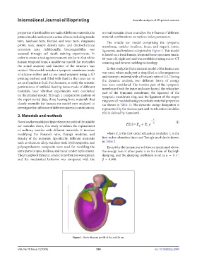
tests, hardness tests, friction and wear tests, roughness The middle ear model comprising the tympanic
profile tests, sample density tests, and electrochemical membrane, ossicles (malleus, incus, and stapes), joints,
corrosion tests. Additionally, biocompatibility was ligaments, and tendons is depicted in Figure 1. This model
assessed through cell death staining experiments. In is based on a fresh human temporal bone specimen (male,
order to create a testing environment similar to that of the 60-year-old, right ear) and was established using micro-CT
human temporal bone, a middle ear model that resembles scanning and reverse molding technology.
the actual anatomy and function of the structure was
created. This model includes a tympanic membrane made In this study, the finite element model of the human ear
of silicone rubber and an ear canal prepared using a 3D was used, where each part is simplified as a homogeneous
printing method and filled with fluid in the inner ear to and isotropic material with a Poisson’s ratio of 0.3. During
the dynamic analysis, two different forms of energy
act as a lymphatic fluid. Furthermore, to verify the acoustic loss were considered. The tension part of the tympanic
performance of artificial hearing bones made of different membrane (both the inner and outer layers), the relaxation
materials, laser vibration experiments were conducted part of the tympanic membrane, the ligament of the
on the printed model. Through a comparative analysis of tympanic membrane ring, and the ligament of the stapes
the experimental data, three hearing bone materials that ring were all modeled using viscoelastic material properties
closely resemble the human ear model were analyzed to (as shown in Table 1). The dynamic energy dissipation is
investigate the influence of different material combinations. represented by the viscous part, and its relaxation modulus
E(t) is defined by Equation I.
2. Materials and methods
Based on the established finite element model of the middle (I)
ear ossicular chain, this study simulates the replacement
of auditory ossicles with different materials. It involves
modifying the Poisson’s ratio, Young’s modulus, and where E is the first-order relaxation modulus; τ is the
1
1
density of the materials. Specifically, different materials first-order relaxation time; and Young’s modulus is shown
such as titanium alloy, stainless steel, hydroxyapatite, and in Table 2.
polycaprolactone composite were used for modeling the Except for the human ear soft tissues mentioned above,
same parts (stapes, malleus, and incus) under replacement. the energy loss of other parts is in the form of Rayleigh
The principle of dynamic conduction vibration was applied, damping, and the damping coefficient is set as α = 0 s ,
-1
and the mechanical behavior was compared with the β = 0.000.
Figure 1. Finite element model of the middle ear.
Volume 10 Issue 3 (2024) 324 doi: 10.36922/ijb.2040