
Page 358 - IJB-10-3
P. 358
International Journal of Bioprinting hNVU chip for brain modeling and drug screening
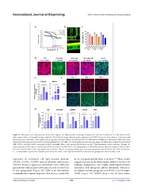
Figure 4. Fabrication and characteristics of the brain region. (A) Measurement of storage modulus (G’) and loss modulus (G”) of the bioink of the
brain region under cooling and heating conditions. (B) Confocal images illustrating the organization of hNVU structures. Red: mixture of pericytes and
astrocytes; green with tube: “brain region”; green (up layer and down layer): endothelial cells. (C) Cell viability of different cell densities within the brain
6
region after printing (the base unit for proportional numbers is 10 cells). (D) Immunofluorescence staining images of cell identity markers for endothelial
cells (CD31), pericytes (NG2), astrocytes (GFAP), microglia (IBA1), and neurons (β3-tubulin) on day 7. Blue represents nuclei. Scale bar: 200 μm. (E)
Gene expression of the neuron markers β3-tubulin and nestin in iPSC-NPCs. (F) Representative immunofluorescence staining images of ECM proteins
(COL4A1 and laminin) in the 7-day experiment. Scale bar: 100 μm. (G) Representative gene expression associated with ECM and cell–ECM interactions.
n = 3, independent experiments. Data are presented as the mean ± S.D. Significant differences between the groups were determined by t-test.
expression of endothelial cell tight junction proteins in the malignant proliferation of glioma. 42,43 These results
(OCLN, CLDN1, CLDN5) and an adhesion link protein suggest that cells in the brain region exhibit a tendency to
(CDH5) showed a significant downward trend, while the undergo angiogenesis and display pathological features
gap junction-related gene GJB6 (gap junction protein beta associated with malignant glioma metastasis. Moreover,
6) was upregulated (Figure 5B). GJB6 is an intercellular we determined the upregulation of SPARC and fibrinogen
communication marker in glioma that plays a crucial role (FGB) (Figure 5A). SPARC plays a role in brain tumor
Volume 10 Issue 3 (2024) 350 doi: 10.36922/ijb.1684