
Page 468 - IJB-10-3
P. 468
International Journal of Bioprinting Biomimetic scaffolds for tendon healing
Concerning the regeneration of partial tendon injuries, exact and comparable way between different structures. It
some interesting results should be highlighted. VEGF165 is expected that this characterization based on modeling
and PDGF-BB had a rapid initial release, favoring the will become a truly relevant tool for the field.
effect of these factors during the first 2 days. Afterward, the
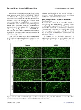
release of VEGF165 still remained fast. This is interesting 3.6.2. In vitro bioactivity of the VEGF165 released
from the scaffold
since the aim of incorporating this factor is to increase the To analyze the activity of the released VEGF165, a
vascularization of the scaffold and the partially ruptured vascularization assay was performed. This assay consisted
tendon mostly during the first days of regeneration (to of analyzing the reorganization of HUVECs on the surface
increase the supply of oxygen and allow the synthesis of the of the scaffold to form tube or capillary-like structures. Two
different types of collagen). In the case of PDGF-BB, after conditions (with and without factor) were tested during
the initial, fast release, the sustained release over time is four sampling times. At each time point, cells were stained
beneficial for proliferation and migration of mesenchymal and the formation of structures was analyzed under the
cells to the damaged tissue. microscope (Figure 9).
These values are widely applied in the pharmaceutical In addition to the qualitative observation, a
field but still not frequently used in tissue engineering field semiquantitative analysis of the area covered by cells, the
to characterize the release profiles from scaffolds in a more total number of loops, the area of all the loops, the mean
Figure 9. In vitro bioactivity of the VEGF165 and formation of pre-vascular structures. Micrographs of the HUVECs on the surface of the scaffolds
(control and with VEGF165) were captured at the different time points at 4×. Scale bar: 500 µm.
Volume 10 Issue 3 (2024) 460 doi: 10.36922/ijb.2632