
Page 470 - IJB-10-3
P. 470
International Journal of Bioprinting Biomimetic scaffolds for tendon healing
the control samples for all time points (Figure 10B, C, research focus of the current work. The concentration was
and E). From the obtained results, it is noteworthy that for selected based on different factors. Higher concentrations
the samples with factor, the number of loops and tubes, as may be appropriate for treatments requiring longer periods
well as their total area and total length, was significantly of time. However, the use of higher concentrations may be
81
greater on days 5 and 9 than other time points (but no subject to greater restrictions imposed by health agencies.
significant differences were detected between day 5 and In this context, lower concentrations (such as 20 ng mL )
-1
day 9). On day 20, however, loops and tubes with a greater would be sufficient to provoke an initial increase of cell
area and length, respectively, were observed in the same proliferation. These cells can then reduce their proliferation
samples (Figure 10D and F). These results demonstrate rate and increase their rate of ECM synthesis.
that the released factor influences HUVECs in vitro under
the tested conditions. 4. Conclusion
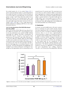
3.6.3. In vitro bioactivity of the PDGF-BB released At present, rest and rehabilitation is the most common
from the scaffold form of treatment for partial tendon injuries. Despite
The in vitro activity of PDGF-BB after being released from the clinical significance due to their prevalent incidence
the scaffold was analyzed with a cell proliferation assay. and the more severe consequences following inadequate
In the cell proliferation assay, control scaffolds (without treatment, there is yet no tissue engineering-based
PDGF-BB) and scaffolds with three concentrations of strategy developed to improve the regeneration process
PDGF-BB (10 ng mL , 20 ng mL , and 50 ng mL ) were of partially injured tendons. Here, we showed that it is
-1
-1
-1
placed on top of a transwell. In the wells of the plate, 2.5 possible to combine multiple biological materials and
× 10 C2C12 cells were cultured. After 24 h, the number 3D bioprinting to obtain a scaffold with a composition
5
of cells in each condition was determined (Figure 11). The similar to that of the tendon ECM. The development
results showed that the factor affects cell proliferation, in of this bioink based on the combination of four
concordance with what has been previously reported in biomolecules (Gel, Alg, HA, and Fg) represents a great
literature indicating that a concentration between 10 and advancement. This bioink possessed good rheological
100 ng mL of PDGF-BB increases cell proliferation and properties necessary for application in 3D bioprinting
-1
viability. Concentrations at 20 ng mL and 50 ng mL were and good degradability and swelling properties.
-1
-1
80
suitable for the regeneration of partial tendon injuries, the Likewise, it also showed good biocompatibility, allowing
Figure 11. Proliferation of C2C12 cells when exposed to different concentrations of PDGF-BB. Results are represented as mean ± SD (n = 3). *p < 0.05.
Volume 10 Issue 3 (2024) 462 doi: 10.36922/ijb.2632