
Page 508 - IJB-10-3
P. 508
International Journal of Bioprinting 3D printing microgroove nerve conduits
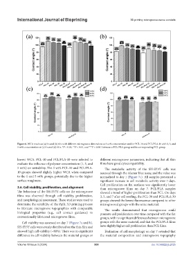
Figure 6. WCA results at (a) 0 s and (b) 60 s with different microgroove dimensions at 3 wt% concentration and for PCL-10 and PCL/PLA-10 with 1, 3, and
5 wt% concentration at (c) 0 s and (d) 60 s. *P < 0.05, **P < 0.01, and ***P < 0.001 between a PCL/PLA group and its corresponding PCL group.
lowest WCA. PCL-10 and PCL/PLA-10 were selected to different microgroove parameters, indicating that all thin
evaluate the influence of polymer concentration (1, 3, and films have good cytocompatibility.
5 wt%) on wettability. The 3 wt% PCL-10 and PCL/PLA- The metabolic activity of the SH-SY5Y cells was
10 groups showed slightly higher WCA when compared assessed through the Alamar blue assay, and the value was
to the 1 and 5 wt% groups, potentially due to the higher normalised to day 1 (Figure 7c). All samples presented a
surface roughness. significant increase in cell metabolic activity over 7 days.
Cell proliferation on flat surfaces was significantly lower
3.6. Cell viability, proliferation, and alignment than microgroove films on day 7. PCL/PLA samples
The behaviour of the SH-SY5Y cells on the microgroove showed a trend of higher proliferation than PCL. On days
films was observed through cell viability, proliferation, 3, 5, and 7 after cell seeding, the PCL-30 and PCL/PLA-30
and morphological assessment. These studies were used to groups showed the lowest fluorescence compared to other
determine the suitability of the PµSL 3D printing process microgrooved groups with the same material.
to fabricate microgroove topographies with comparable The results demonstrated that microgrooves could
biological properties (e.g., cell contact guidance) to promote cell proliferation over time compared with the flat
conventionally fabricated microgroove films. groups, with no significant differences between microgroove
Cell viability was assessed on day 7 (Figure 7a and b). groups with the same material, and that the PCL/PLA films
SH-SY5Y cells were evenly distributed on the thin film and have slightly higher cell proliferation than PCL films.
showed high cell viability (~80%). There was no significant Evaluation of cell morphology on day 7 revealed that
difference in cell viability between the material groups or the material composition and microgroove topography
Volume 10 Issue 3 (2024) 500 doi: 10.36922/ijb.2725