
Page 50 - IJB-4-1
P. 50
Jang T-S, et al.
scaffolds in tension and compression tests significantly
increased with the addition of Laponite XLG. The release
of silicon and magnesium ions from Laponite XLG
promoted the proliferation and differentiation of primary rat
osteoblast (ROB) cells. PNAGA-clay hydrogel scaffolds
implanted in tibia defects of rats effectively induced new
bone formation.
The possibilities of bioprinting and growth factor
delivery of hydrogel-nanoclay composites were verified
by Ahlfeld et al. [98] Laponite XLG was blended with various
compositions of alginate-methylcellulose hydrogels. The
pastes were printed by 3D plotting method and incubated
in CaCl solution. Human telomerase reverse transcriptase-
2
mesenchymal stem cells (hTERT-MSC) were mixed with
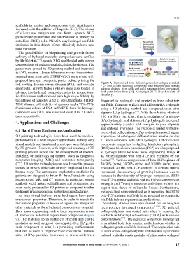
prepared hydrogel composite pastes before printing for Figure 9. Customized bone defect regeneration using a extruded
cell plotting. Bovine serum albumin (BSA) and vascular PLA and gelatin hydrogel composite with incorporated human
endothelial growth factor (VEGF) were also loaded in adipose derived stem cells and gold nanoparticles (reproduced
advance into hydrogel composite pastes for release tests. with permission from [89]. Copyright 2017, Royal society of
chemistry).
Scaffolds were well-extruded with high shape fidelity by
the addition of nanoclay. After 21 days, the printed hTERT- dispersed in hydrogels and printed as bone substitute
MSC showed cell viability of approximately 70%–75%. scaffolds. Demirtas et al. printed chitosan-HA hydrogels
Continuous release of BSA and VEGF, from the hydrogel using a 3D plotting method and compared them with
composite scaffolds, was observed even after 21 and 7 alginate-HAp hydrogels [104] . With the addition of about
days, respectively. 180 nm HAp particles, elastic modulus of alginate-
4. Applications and Challenges HAp hydrogels and chitosan-HAp hydrogels increased
approximately 3-and-5 fold compare to pure alginate
and chitosan hydrogels. The hydrogels loaded with pre-
4.1 Hard Tissue Engineering Application oseteoblast cells, chitosan-HAp hydrogels showed higher
3D printing technologies have been used by medical expression of osteogenic differentiation marker on day
professionals in a wide range of applications. Initially, only 21 when compared with other hydrogels. Other calcium
visual models and functional prototypes were fabricated phosphate materials including bicalcium phosphate
by 3D printers. However, with improved accuracy of 3D (BCP) and tricalcium phosphate (TCP) are also proposed
printing process as well as the development of medical as hydrogel fillers for bone tissue engineering. Diogo et
imaging, or radiology equipment such as magnetic al. mixed alginate with beta-TCP and extruded by 3D
resonance imaging (MRI) and computed tomography plotter [105] . Various composition of beta-TCP/alginate of
(CT), 3D printing technologies can now be used to produce 50/50% (w/w), 30/70% (w/w) and 20/80% (w/w) were
tissues or organs which are directly implanted into the evaluated. As the beta-TCP contents in alginate matrix
human body. The customized implantable scaffolds for increased, the accuracy of printing increased due to
patients are designed to better fit the affected site using increase in the viscosity of hydrogel composites. 50/50
reconstructed MRI and CT images. In particular, porous beta-TCP/alginate scaffolds had the highest compression
scaffolds which induce cell infiltration and proliferation are strength and Young’s modulus and these values are
more easily produced by 3D printers as compared to other higher than those of trabecular bones. Furthermore,
traditional processes such as subtractive manufacturing. biological test using osteoblast cells suggested that 50/50
As mentioned before, pure hydrogels have poor beta-TCP/alginate scaffolds have potential as composite
mechanical properties. Therefore, in order to match the scaffolds in bone regeneration applications.
mechanical properties of tissues or organs, the integration Similarly, studies were also carried out on bioglass
other materials to form hydrogel composites is essential. incorporated hydrogel composites [106] . 3D printed
Hard tissue engineering such as bone regeneration is one collagen/alginate was coated with silica by soaking the
of biomedical fields that require these composites (Figure scaffolds in tetraethyl orthosilicate (TEOS) with various
9). The material needs sufficient strength and elastic concentrations [107] . The scaffolds were more mineralized
modulus as well as good biocompatibility. HAp, the in simulated body fluid solution as the fractions of silica in
main component of bone, is a promising reinforcement collagen/alginate scaffolds increased. The degradation rate
that can be used to improve these conditions. Various of silica coated collagen/alginate scaffolds was significantly
sizes of HAp particles from nano to micro scale were reduced while the elastic modulus of silica coated collagen/
International Journal of Bioprinting (2018)–Volume 4, Issue 1 17