
Page 52 - IJB-4-1
P. 52
Jang T-S, et al.
al. [113] The addition of alginate not only improved the hydrogel and piezoelectric inkjet, cell damage mainly results from
viscosity and shape fidelity, but also increased the tensile the thermal heating during the printing process, whereas in
strength and toughness of hydrogels. extrusion bioprinting, compression forces and shear stresses
The skin is the largest organ that covers the human body and generated during the printing causes damage to cells [120] .
it plays an important role in regulating temperature, controlling On the other hand, biocompatible hydrogels widely used
evaporation as well as protecting from pathogens and external for matrix materials of cell-laden bioinks or supporting
environment. It is a complex structure with three sequential materials of printed cells require solidification strategies,
layers including epidermis which is the outer layer, dermis e.g., photo-crosslinking, in situ chemical crosslinking,
that is permeated by a complex nervous and blood vessel, and physical crosslinking or shear-thinning [121-124] . Integration
hypodermis consisting of subcutaneous tissue [114] . Therefore, in of those solidification methods into bioinks is challenging,
skin tissue engineering, many researchers tried to substitute this particularly in case of cell-laden hydrogel bioinks where
complex and important organs with artificial skin grafts such the hydrogel gelation process should minimize the potential
as hydrogels for curing skin wounds and diseases [115] . With damage of encapsulated cells [121–123,125,126] . Particularly,
recent advances in hydrogel printing technique which moved UV-based photopolymerization reactions of bioactive
from 2D to 3D printing allow more flexibility in controlling hydrogels (e.g., gelatin, collagen, chitosan) are commonly
the micro/nano level structure. Moreover, studies are focused coupled with bioprinting, employed either during the
[39]
on 3D printing hydrogel composites to functionalize hydrogel printing process [127] or after the deposition of bioinks to
scaffolds that are closely mimicking real skin tissue. produce stable 3D hydrogels with intricate architectures
Skardal et al. investigated the possibility of skin for cell encapsulation. However, the deleterious effects of
regeneration of mouse skin wound by printed amniotic UV light irradiation and cytotoxicity of radicals generated
fluid-derived stem (AFS) cells incorporated hydrogels [116] . by photoinitiators lead to a decrease in cell viability and
They used fibrinogen/collagen mixed with 50:50 volume ultimately DNA damage [128] .
ratio as hydrogel composites and hydrogel composites 4.3 Vascular Application
including AFS cells and mesenchymal stem cells (MSCs).
Fibrinogen/collagen hydrogel composites with cells and Fabrication of vascular system is one of the main
thrombin were directly printed on the skin wound of nude challenges in 3D printing, because isolated cells cannot
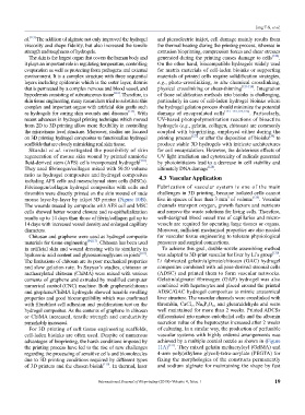
3
mouse layer-by-layer by inkjet 3D printer (Figure 10B). live in spaces of less than 3 mm of volume [129] . Vascular
The wounds treated by composite with AFS cell and MSC channels transport oxygen, growth factors and nutrients
cells showed better wound closure and re-epithelialization and remove the waste solutions for living cells. Therefore,
results up to 14 days than those of fibrin/collagen gel up to well-designed blood vessel tree of capillaries and micro-
14 days with increased vessel density and enlarged capillary vessels are required for operating large tissues or organs.
diameters. Moreover, sufficient mechanical properties are also needed
Chitosan and graphene were used as hydrogel composite for vascular tissue engineering to tolerate physiological
materials for tissue engineering [84,117] . Chitosan has been used pressures and surgical connections.
in artificial skin and wound dressing with its similarity in To achieve this goal, double-nozzle assembling method
hyaluronic acid content and glycosaminoglycans in joints [118] . was adapted to 3D-print vascular for liver by Li’s group [130] .
The limitations of chitosan are its poor mechanical properties Li fabricated gelatin/alginate/chitosan (GAC) hydrogel
and slow gelation rate. In Sayyar’s studies, chitosan or composites combined with adipose-derived stromal cells
methacrylated chitosan (ChiMA) were mixed with various (ADSC) and printed them to form vascular networks.
contents of graphene and extruded by modified computer Gelatin/alginate/ fibrinogen (GAF) hydrogel was also
numerical control (CNC) machine. Both graphene/chitosan combined with hepatocytes and placed around the printed
and graphene/ChiMA hydrogels showed tunable swelling ADSC/GAC hydrogel composites to mimic anatomical
properties and good biocompatibility which was confirmed liver structure. The vascular channels were crosslinked with
with fibroblast cell adhesion and proliferation test on the thrombin, CaCl , Na P O and glutaraldehyde and were
5 3
2
10
hydrogel composites. As the contents of graphene in chitosan well maintained for more than 2 weeks. Printed ADCSs
or ChiMA increased, tensile strength and conductivity differentiated into mature endothelial cells and the albumin
remarkably increased. secretion value of the hepatocytes increased after 2 weeks
For 3D printing of soft tissue engineering scaffolds, of culturing. In a similar way, the production of perfusable
cell-laden bioinks are often used. Despite of numerous vascular systems with highly ordered arrangements was
advantages of bioprinting, the harsh conditions imposed by achieved by a multiple coaxial nozzle as shown in (Figure
the printing process have led to the rise of new challenges 11A) [131] . They mixed gelatin methacryloyl (GelMA) and
regarding the processing of sensitive cells and biomolecules 4-arm poly(ethylene glycol)-tetra-acrylate (PEGTA) for
due to 3D printing conditions required by different types fixing the morphologies of the constructs permanently
of 3D printers and the chosen bioink [119] . In thermal, laser and sodium alginate for maintaining the shape by fast
International Journal of Bioprinting (2018)–Volume 4, Issue 1 19