
Page 40 - IJB-4-2
P. 40
Bioprinting of artificial blood vessels
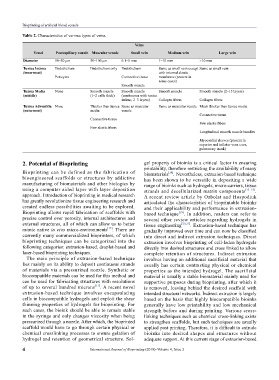
table 2. Characteristics of various types of veins.
veins
vessel Postcapillary venule Muscular venule Small vein Medium vein large vein
Diameter 10–50 μm 50–100 μm 0.1–1 mm 1–10 mm >10 mm
tunica intima Endothelium Endothelium only Endothelium Same as small vein except Same as small vein
(innermost) with internal elastic
Pericytes Connective tissue membrane (present in
some cases)
Smooth muscle
tunica Media None Smooth muscle Smooth muscle Smooth muscle Smooth muscle (2–15 layers)
(middle) (1–2 cells thick) (continuous with tunica
intima; 2–3 layers) Collagen fibers Collagen fibers
tunica Adventitia None Thicker than tunica Same as muscular Same as muscular venule Much thicker than tunica media
(outermost) media venule
Connective tissue
Connective tissue
Few elastic fibers
Few elastic fibers
Longitudinal smooth muscle bundles
Myocardial sleeves (present in
superior and inferior vena cava,
pulmonary trunk)
2. Potential of Bioprinting gel property of bioinks is a critical factor in ensuring
printability, therefore restricting the availability of many
Bioprinting can be defined as the fabrication of biomaterials . Nevertheless, extrusion-based technique
[14]
bioengineered scaffolds or structures by addictive has been shown to be versatile in depositing a wide
manufacturing of biomaterials and other biologics by range of bioinks such as hydrogels, micro-carriers, tissue
using a computer aided layer with layer deposition strands and decellularized matrix components [15–18] .
approach. Introduction of bioprinting in medical research A recent review article by Ozbolat and Hospodiuk
has greatly revolutionize tissue engineering research and articulated the characteristics of bioprintable bioinks
created endless possibilities awaiting to be explored. and their applicability and performance in extrusion-
Bioprinting allows rapid fabrication of scaffolds with based technique [19] . In addition, readers can refer to
precise control over porosity, internal architectures and several other review articles regarding hydrogels in
external structures, all of which can allow us to better tissue engineering [20,21] . Extrusion-based technique has
mimic native in vivo micro-environments [13] . There are gradually improved over time and can now be classified
currently many commercialized bioprinters, of which into direct and indirect extrusion techniques. Direct
bioprinting techniques can be categorized into the extrusion involves bioprinting of cell-laden hydrogels
following categories: extrusion-based, droplet-based and directly into desired structures and cross linked to allow
laser-based bioprinting techniques. complete retention of structures. Indirect extrusion
The main principle of extrusion-based technique involves having an additional sacrificial material that
lies mainly on its ability to deposit continuous strands usually has certain contrasting physical or chemical
of materials via a pressurized nozzle. Synthetic or properties as the intended hydrogel. The sacrificial
biocompatible materials can be used for this method and material is usually a stable biomaterial mainly used for
can be used for fabricating structures with resolutions supportive purposes during bioprinting, after which it
of up to several hundred microns [11] . A recent novel is removed, leaving behind the desired scaffold with
extrusion-based technique involves encapsulating intended structural networks. Indirect extrusion is largely
cells in biocompatible hydrogels and exploit the shear based on the basis that highly biocompatible bioinks
thinning properties of hydrogels for bioprinting. For generally have low printability and low mechanical
such cases, the bioink should be able to remain stable strength before and during printing. Various cross-
in the syringe and only changes viscosity when being linking techniques such as chemical cross-linking exists
pressurized through a nozzle. After which, the bioprinted to strengthen scaffolds, but such techniques are usually
scaffold would have to go through certain physical or applied post printing. Therefore, it is difficult to extrude
chemical crosslinking processes to ensure gelation of bioinks into desired shapes and structures without
hydrogel and retention of geometrical structure. Sol- adequate support. At this current stage of extrusion-based
4 International Journal of Bioprinting (2018)–Volume 4, Issue 2