
Page 42 - IJB-4-2
P. 42
Bioprinting of artificial blood vessels
might be due to the fact that UV crosslinking requires made it a popular choice for fabrication of both vascular
only about 8 s per layer but visible light crosslinking graft and vascularized tissue constructs. There are
system takes about 4 min per layer. However, this novel currently three major approaches for fabrication of blood
technique was proposed to be safer for maintaining vessels: perfusable scaffolds, self-assembly of vessels
long-term cell functionality due to the absence of UV. and scaffold free biofabrication of autonomous vascular
A recent extensive parametric study done by Koch L, structures.
et al. investigated the effects and inter-connectivity of 2.1 Perfusable Scaffolds
laser wavelength, pulse duration, pulse energy, focal
spot size and laser intensity on different parameters such During the last decade, a lot of emphasis has been
[32]
as droplet volumes and cell viability . It was reported placed on developing 3D models to attempt to mimic in-
that with shorter wavelengths, less energy was required vivo native tissue micro-environment. In general, 3D
to print a certain droplet volume. However, this effect culture systems are an assembly of different cell lines
can be compensated by increasing laser pulse energy. and primary cells into a 3D model with either scaffolds
In short, there was no optimal and best parameters for or cell culture plates [42] . There are several existing 3D
laser-based technique and that other parameters such as models currently available and there are some models
long-term stability or inexpensiveness can be used on being developed to model our human organs. Even
case-to-case basis consideration for each study. Although though each 3D model has its own advantages and
various techniques can be used in fabrication of vascular disadvantages, generally all 3D models are found to
grafts or vascularized constructs, each technique varies have more similarity to native organs compared to 2D
[43]
considerably in generating different types of desired cultures . Therefore, 3D models are deemed as the way
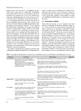
constructs. Table 3 provides a general overview of the ahead for personalized medicine, drug screening and
advantages and disadvantages of the above mentioned physiological studies of diseases. However, even though
methods in vascular engineering. such 3D models and arrangements enable cell-cell
The ability of bioprinting to generate constructs with interactions similar to a native-like micro-environment,
well-defined architectures and precise cell deposition has there remains a need for vascularization within the organ
table 3. Advantages and disadvantages of extrusion-based, droplet-based and laser-based technique in bioprinting of blood vessels
techniques Advantages Disadvantages References
extrusion-based Direct extrusion-based bioprinting allows Direct extrusion-based: challenging to bioprint bifurcated vessels. [33–35]
fabrication of large scaffolds with high aspect
ratios high aspect ratio. Indirect extrusion-based: fixed height for each layer thus limiting
Indirect extrusion-based bioprinting allows height of sacrificial ink or lumen. Increasing height of sacrificial ink
fabrication of highly complex patterns with alone might cause occlusion of lumen when removed.
sacrificial ink which can be removed to generate
lumens for perfusion. Current studies have only managed to line lumen with single layers
of endothelial cells and is unable to mimic the various layers in native
vessels.
Most studies use hydrogels as their main material which tend to
degrade over time thus causing collapse of lumen.
Studies have shown that cells need to be deposited in certain patterns
for efficient vascularization which is something unachievable with
extrusion-based technique due to its lack of specificity in cells seeding.
Droplet-based It allows for fabrication of high-resolution Unable to fabricate large constructs. [36–38]
constructs with co-culture of multiple cell types.
Hydrogels with high viscosity of above 0.1 Pa.s-1 easily clogs up
It allows on-demand ejection of very small nozzle causing low reproducibility, high shear stress and unwanted
droplets of bioink. cellular aggregation. This greatly limits the choice of hydrogels for
droplet-based bioprinting.
laser-based It allows for fabrication of constructs with well- Unable to achieve sufficient cell density as compared to native [39–41]
defined structures with bifurcated branches. composition.
Almost zero shear stress related damages to cells Requirement of photo-curable bioink limits the choice of available
as cells are not ejected via nozzles. Thus cell bioink. Photo-initiators are toxic in general which can cause unwanted
viability is significantly higher when compared to cellular damage.
other available techniques.
Effects of constant and prolonged laser irradiation on cells are not yet
It can be used to print hydrogels with high established.
viscosity.
6 International Journal of Bioprinting (2018)–Volume 4, Issue 2