
Page 10 - IJB-10-4
P. 10
International Journal of Bioprinting PAI for 3D bioprinted constructs
towards complex structural hierarchies and larger- emerge as powerful tools, offering sub-micrometer spatial
scale volumes (Figure 1). 15–17 Bioprinting is a bottom- resolution to discern intricate 3D microarchitectures and
up fabrication process that begins at the cellular and unveil the nuances of complex cellular physiology through
material levels to create constructs with a high hierarchical labeling. Techniques such as confocal microscopy, 22,23
complexity, such as tissues and organs. As this hierarchy light sheet microscopy, 24–26 and optical coherence
evolves, the physical scale spans from the micrometer to tomography, 27–29 have played instrumental roles in
the centimeter scale, prompting exploration into various advancing tissue engineering. However, these techniques
key parameters. These parameters encompass the have limitations in imaging biological structures thicker
18
chemical, mechanical, and biocompatibility attributes of than the optical diffusion limit (<1 mm), owing to the
printable materials at the microarchitectural level; cellular opaque nature of biological tissue. Consequently, imaging
orientation and functional maturation at the tissue level; thicker tissues necessitates destructive sample preparation
and structural morphology and systematic function at the process, such as sectioning or tissue clearance, which can
organ level. 19–21 compromise the integrity of the printed tissue.
Bioimaging serves as a crucial method for inspecting In contrast, photoacoustic imaging (PAI) stands out
accurate tissue formulations in terms of their structural for its exceptional imaging depth, owing to its acoustic
morphology and biological development, including hybridity. 30–35 PAI conjugates the photoacoustic (PA)
cell viability, differentiation, and complex physiological effect, wherein chromophores emit acoustic waves upon
pathways. In this domain, 3D optical imaging techniques instant thermal expansion following nanosecond pulse
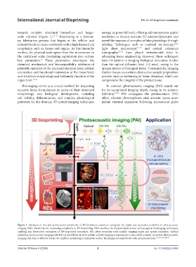
Figure 1. Advances in size and architectural complexity of 3D-bioprinted constructs alongside the depth and resolution scalability in photoacoustic
imaging (PAI). Parallel to the increasing complexity in 3D bioprinting, PAI visualizes the deepest depth across various optical bioimaging techniques,
enabling non-destructive evaluation of 3D-bioprinted constructs. PAI offers versatility with scalable imaging depth and spatial resolution. Optical
resolution photoacoustic imaging (OR-PAI) is suitable for in vitro cellular activity imaging at micrometer scales, while acoustic resolution photoacoustic
imaging (AR-PAI) is effective for in vivo implant monitoring at millimeter scales. The images are reproduced with permission from. 19, 21, 45, 50, and 59
Volume 10 Issue 4 (2024) 2 doi: 10.36922/ijb.3448