
Page 12 - IJB-10-4
P. 12
International Journal of Bioprinting PAI for 3D bioprinted constructs
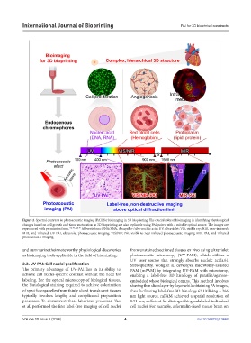
Figure 2. Spectral contrasts in photoacoustic imaging (PAI) for bioimaging in 3D bioprinting. The crucial roles of bioimaging in identifying physiological
changes based on cell growth and tissue maturation in 3D bioprinting are also replicable using PAI paired with a suitable optical source. The images are
reproduced with permission from. 64, 70, and 79 Abbreviations: DNA/RNA, deoxyribo-/ribo-nucleic acid; UV, ultraviolet; VIS, visible ray; NIR, near-infrared;
MIR, mid-infrared; UV-PAI, ultraviolet-photoacoustic imaging; VIS/NIR-PAI, visible to near-infrared photoacoustic imaging; MIR-PAI, mid-infrared
photoacoustic imaging.
and summarize their noteworthy physiological discoveries from unstained sectioned tissues ex vivo using ultraviolet
as bioimaging tools applicable in the field of bioprinting. photoacoustic microscopy (UV-PAM), which utilizes a
UV laser source that strongly absorbs nucleic acids.61
2.2. UV-PAI: Cell nuclei proliferation Subsequently, Wong et al. developed microtomy-assisted
The primary advantage of UV-PAI lies in its ability to PAM (mPAM) by integrating UV-PAM with microtomy,
achieve cell nuclei-specific contrast without the need for enabling a label-free 3D histology of paraffin/agarose-
labeling. For the optical microscopy of biological tissues, embedded whole biological organs. This method involves
the histological staining required to achieve colorization shaving thin slices layer-by-layer while obtaining PA images,
of specific organelles from thinly sliced translucent tissues thus facilitating label-free 3D histology.62 Utilizing a 266
typically involves lengthy and complicated preparation nm light source, mPAM achieved a spatial resolution of
processes. To circumvent these laborious processes, Yao 0.91 μm, sufficient for distinguishing unlabeled individual
et al. performed the first label-free imaging of cell nuclei cell nuclei. For example, a formalin-fixed mouse brain on
Volume 10 Issue 4 (2024) 4 doi: 10.36922/ijb.3448