
Page 16 - IJB-10-4
P. 16
International Journal of Bioprinting PAI for 3D bioprinted constructs
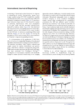
monitoring of functional cerebral dynamics in response approaches remains challenging. A deeply seated placenta
to physiological stimuli. Demonstrative mouse brain is normally inaccessible with conventional optical imaging
images acquired using UFF-PAM revealed the complete techniques. Biomedical tomography such as magnetic
vascular network of the cortex, along with maps of the sO2 resonance imaging or X-ray computed tomography
delineated by individual vessels (Figure 6a). A maximum require contrast agent administration for angiographic
rapid volumetric frame rate of 2 Hz allowed the capture presentation but still lack sufficient spatial resolution
of spreading depolarization (SD) wave propagation and molecular sensitivity. The advent of UFF-PAM via
74
in the gray matter and the associated hemodynamics placental window overcomes these technical limitations, as
induced by ischemic stroke (Figure 6b). This facilitated being a high-resolution, high-speed, in-depth functional
the measurement of a propagating speed of 2.56 mm/min, microvessel imaging technique providing intrinsic optical
alongside a precise determination of the origin location contrast from hemoglobin. They longitudinally monitored
for each SD wave. A continuous monitoring of the vessel the daily changes in the mouse placenta during a healthy
diameter (Figure 6c) and sO2 (Figure 6d) during the pregnancy term, from 7 to 19 embryonic days. Moreover,
propagation of SD waves revealed local vasoconstriction the observed changes in each parameter displayed notable
and hypoxia in response to spreading ischemia. correlations with the specific trimesters of pregnancy. Rapid
In a demonstration of the application of UFF-PAM, microvessel development was evident in the first trimester,
Zhu et al. achieved a longitudinal imaging of the placental whereas the second trimester witnessed an increase in both
vasculature of maternal mouse using an intravital window. the quantity and size of placental vessels. Throughout the
71
The placental vasculature, which supplies nutrients and third trimester, vascular development persisted along with
oxygen crucial for embryo development and growth, elevated vascular branching. Notably, sO levels reflect the
2
plays a vital role in embryonic development and growth. transition from indirect oxygen delivery via trophoblasts
While microvascular alterations and placental pathologies to direct delivery via fetal circulation.
have been correlated from histological findings, 72,73 Rebling et al. utilized large-scale optoacoustic
investigation of hemodynamic functions and metabolic microscopy (LSOM) to longitudinally image the
changes in mouse placental vessels using in vivo imaging revascularization process during wound healing, thereby
Figure 6. Ultrafast functional photoacoustic microscopy (UFF-PAM) presenting pathophysiological changes in vasculature. (a) A mouse brain image over
the entire cortex and its corresponding topical oxygen saturation (sO2) map measured from 532/558 nm dual-wavelength excitation. (b) Propagation time
map of spreading depolarization (SD) waves observed from a stroke-induced mouse model. (c) Monitoring of vessel diameter changes and (d) change in
sO2 during SD wave propagation. The images are reproduced with permission from. 70
Volume 10 Issue 4 (2024) 8 doi: 10.36922/ijb.3448